High dose chemotherapy and autologous hematopoietic stem cell rescue for children with high risk neuroblastoma: Two case report
Introduction: High-risk neuroblastoma is a childhood malignancy with a poor prognosis. Gradual
improvements in survival have correlated with therapeutic intensity, and the ability to harvest, process and
store autologous hematopoietic stem cells has allowed for dose intensification beyond marrow tolerance.
Case: We report two cases which were diagnosed high risk neuroblastoma. The first case had big
heterogeneous at his left adrenal gland with metastasis in bone marrow and his brain. The second case
with localised tumor at her right kidney and NMYC positive. Both cases were treated with SIOPEN protocol,
starting with 8 course RAPID COJECT. Patients had partial response and were collected stem cells. Then
they were primary tumor resection; receiving high dose chemotherapy with autologous- stem cell rescue,
radiation and retino acid treatment, respectively.
Conclusion: This is initially report of autologous stem cell transpant for high risk neuroblastoma. Even
only two patients, the results showed that stem cell transplant was safe and effective. We are continuing
to implement for more case with high risk neuroblastoma and will have better report with a bigger number.
Trang 1
Trang 2
Trang 3
Trang 4
Trang 5
Tóm tắt nội dung tài liệu: High dose chemotherapy and autologous hematopoietic stem cell rescue for children with high risk neuroblastoma: Two case report
Bệnh viện Trung ương Huế
94 Journal of Clinical Medicine - No. 62/2020
HIGH DOSE CHEMOTHERAPY AND AUTOLOGOUS
HEMATOPOIETIC STEM CELL RESCUE FOR CHILDREN
WITH HIGH RISK NEUROBLASTOMA: TWO CASE REPORT
Nguyen Thi Kim Hoa1, Tran Kiem Hao1, Chau Van Ha1, Phan Canh Duy2,
Dong Si Sang3, Phan Thi Thuy Hoa3, Ho Huu Thien4, Nguyen Thi My Linh1, Pham Nhu Hiep4
DOI: 10.38103/jcmhch.2020.62.16
ABSTRACT
Introduction: High-risk neuroblastoma is a childhood malignancy with a poor prognosis. Gradual
improvements in survival have correlated with therapeutic intensity, and the ability to harvest, process and
store autologous hematopoietic stem cells has allowed for dose intensification beyond marrow tolerance.
Case: We report two cases which were diagnosed high risk neuroblastoma. The first case had big
heterogeneous at his left adrenal gland with metastasis in bone marrow and his brain. The second case
with localised tumor at her right kidney and NMYC positive. Both cases were treated with SIOPEN protocol,
starting with 8 course RAPID COJECT. Patients had partial response and were collected stem cells. Then
they were primary tumor resection; receiving high dose chemotherapy with autologous- stem cell rescue,
radiation and retino acid treatment, respectively.
Conclusion: This is initially report of autologous stem cell transpant for high risk neuroblastoma. Even
only two patients, the results showed that stem cell transplant was safe and effective. We are continuing
to implement for more case with high risk neuroblastoma and will have better report with a bigger number.
 1. Pediatric Center, Hue Central Hospital
 2. Oncology Center, Hue Central Hospital
 3. Blood and Transfusion Center, Hue Central Hospital
 4. Department of Abdominal Emergency and Pediatric
Surgery, Hue Central Hospital
Corresponding author: Nguyen Thi Kim Hoa
Email: kimhoa.fmi@gmail.com
Received: 8/5/2020; Revised: 17/5/2020
Accepted: 20/6/2020
I. INTRODUCTION
Neuroblastoma is the most common extracranial
solid tumor in children[1], accounting for 7% to 8%
of all childhood cancers. The prevalence is about 1
case per 7.000 live births. Neuroblastoma is slightly
more common in boys than in girls, with a male to
female ratio of 1.1 to 1. The peak incidence occurs
at 2 years of age, some studies show that 89% are
younger than 5 years, and 98% of cases are diagnosed
in the first 10 years of life. The distribution of cases
by age clearly shows that this is a disease of infancy
and early childhood, with the highest number of
cases diagnosed in the first month of life. Before
the development of comprehensive, multimodality
therapy, long term survival probabilities for high
risk neuroblastoma patient were less than 15% [2].
High dose chemotherpay with autologous stem
cell rescue has improved the outcomes for children
with high risk neuroblastoma [3,4]. Using busulfan
and melphalan (BuMel) as conditioning regime,
the International Society of Pediatric Oncology
Neuroblastoma HR NBL1 protocol reported 3 year
High dose chemotherapy and autologous...
Hue Central Hospital
Journal of Clinical Medicine - No. 62/2020 95
EFS and OS of 49% and 60%, respectively[2].
Since 2016, Hue Pediatric Center has been starting
to treat for children with solid tumor, and has been
receiving many neuroblastoma patients, in which
there are 60% high risk neuroblastoma. How to
improve the survival rate for high risk neuroblastoma
is a challenge for our team. With the great support
from Board Director of Hue Central Hospital, Hue
Pediatric Center did autologous transplant sucessfully
for two high risk neuroblastoma cases, and hopefully
we has been continuing to do more cases in order
to build Hue Central Hospital as one of Vietnam’s
leading medical center.
II. CASE REPORT
2.1. Clinical features and imaging finding
The first case: 5 year old male was admitted
to Hue Pediatric Center due to fever and pain at
his right knee. He was done CT scan whole body
and found out one heterogeneous tumor at his
retroperitoneum, measuring 8.6x5.1x4.9cm, with
calcification inside. The tumor encased some
arteries at retroperitoneum, invased left kidney and
left adrenal gland. At his occipital area and sinciput
area, there were lesions measuring from 10-40 mm.
He was done bilateral bone marrow biopsy and the
result showed metastasis neuroblastoma.
The second case: 5 year old female was admitted to
Hue Pediatric Center due to She came to my hospital
due to her abdominal distention. She was done CT
scan whole body and found out one heterogeneous
tumor at her right kidney measuring 7.9x8.3x8.9 cm
(Figure 1, Figure 2). This tumor compressed right
kidney and the right kidney had hydronephrosis. The
tumor also invased the liver and right adrenal gland.
She was done bilateral bone marrow biopsy and the
result was negative. She was done needle biopsy with
the result: neuroblastoma, NMYC: positive.
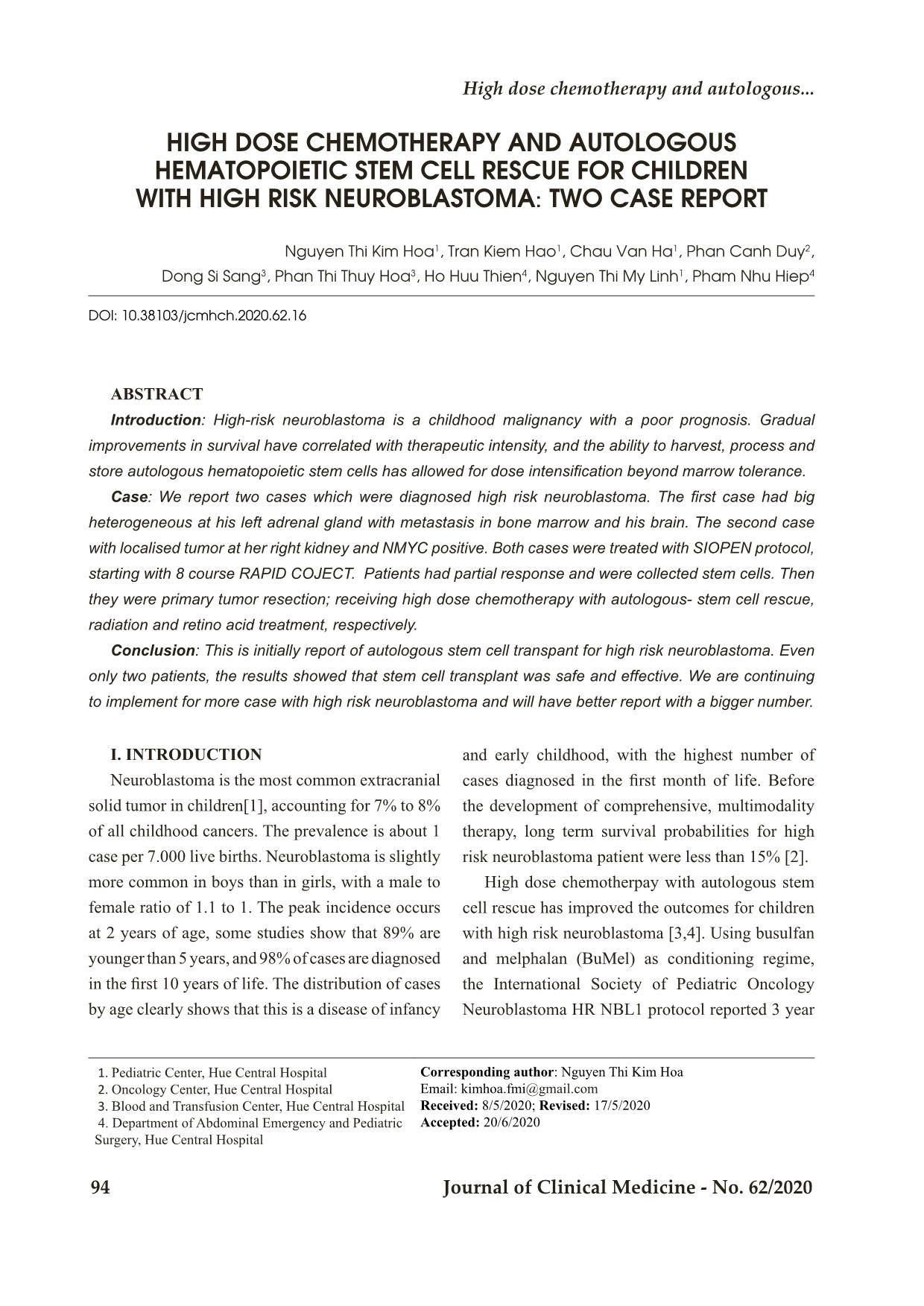
Figure 1: CT scan whole body of the first patient showed tumours at his abdomen and his brain (at initial time)
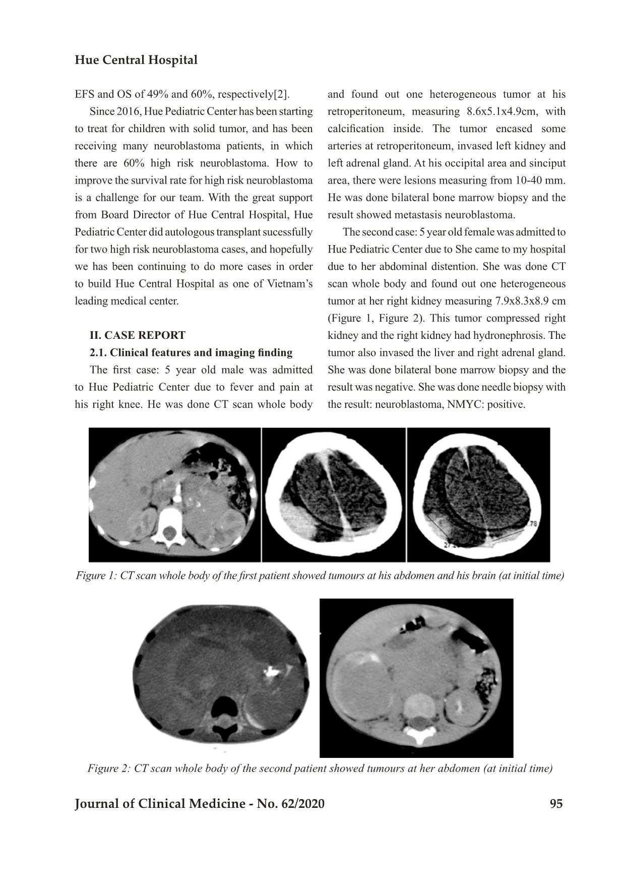
Figure 2: CT scan whole body of the second patient showed tumours at her abdomen (at initial time)
Bệnh viện Trung ương Huế
96 Journal of Clinical Medicine - No. 62/2020
2.2. Pathology results
For the first case, we did needle biopsy the
primary tumor and the result showed neuroblastoma,
with synaptophysin, chromogranin: positive, LCA,
CK AE1/3, Vimemtin, CD 99: negative. The result
of bilateral bone marrow biopsy were reviewed in St
Jude Children’s Research Hospital. The result showed
that the mononuclear cells were subset positive for
vimentin, chromogranin A, synaptophysin, CD
99dim while negative for keratin AE1/3, EMA. CD
43, myogenin, MyoD1, CD42b. These finding were
consistent with metastatic neuroblastoma.
After remove the residual tumor, we sent the
sample to St Jude Children’s Research Hospital
in Memphis to review. The result showed:
microscopic examination of the slide showed a
large nerve fascicle attached to a small fragment
of the tumor with evidence of therapy effect. The
tumor was composed of Schwannian stroma with
scattered neuroblastic cells in different stages of
maturation along with ganglion cells and scattered
hemosiderin - laden macrophages. Fuorescence in
situ hybridization for MYNC performed at St. Jude
was negative for amplification.
The result of second case:
The histologic sections showed a neuroblastic
tumor composed of sheets of malignant small round
cells with minimal interspersed fibrillary neuropil-
like material and large areas of tumor necrosis.
The tumor cells exhibitted brisk mitosis with
karyorrhexis in more than 4% of the tumor cells.
Immunohistochemically, the tumor cells showed
patchy positive staining for synaptophysin, whereas,
cytokeratine AE1/AE3, desmin, BCOR, and CD 99
are negative. The overall features were consistent
with a poorly differentiated neuroblastoma with an
unfavorable histopathology.
Fluorescence in situ hybridization testing
performed at St. Jude showed MYCN amplification.
2.3. Diagnosis and treatment and evaluation
Both cases were diagnosed high risk neuroblas-
toma and were treated with SIOPEN protocol for
high risk neuroblastoma. They were received 8
course of RAPID COJECT.
After 8 course chemotherapy, both patients had partial respones.
The first patient: the tumor at his adrenal gland shrank significantly, with the dimesion: 3.6x3.1x2.8 cm.
The lesion at his brain membrane at the right side of sinciput shrank, measuing: 39x10 mm (Figure 3,
Figure 4).
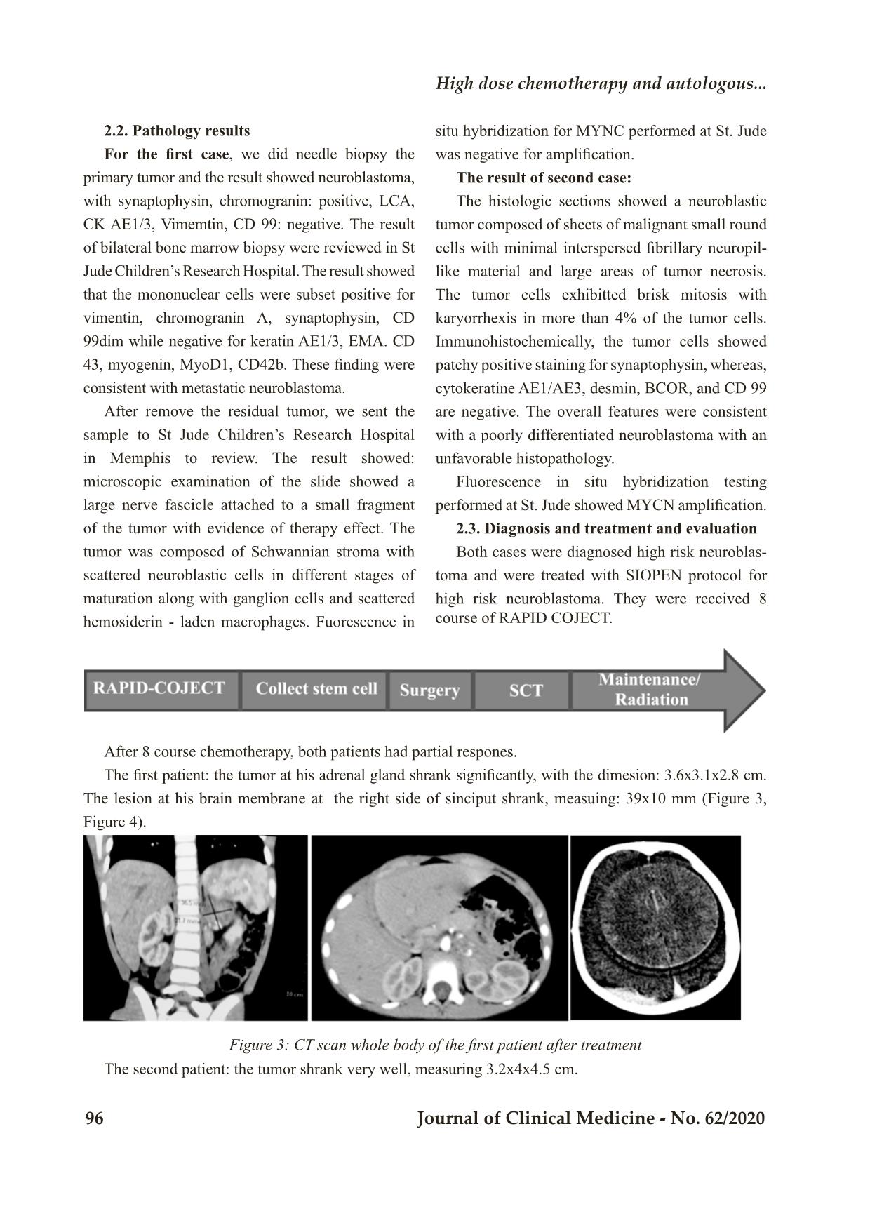
Figure 3: CT scan whole body of the first patient after treatment
The second patient: the tumor shrank very well, measuring 3.2x4x4.5 cm.
High dose chemotherapy and autologous...
Hue Central Hospital
Journal of Clinical Medicine - No. 62/2020 97
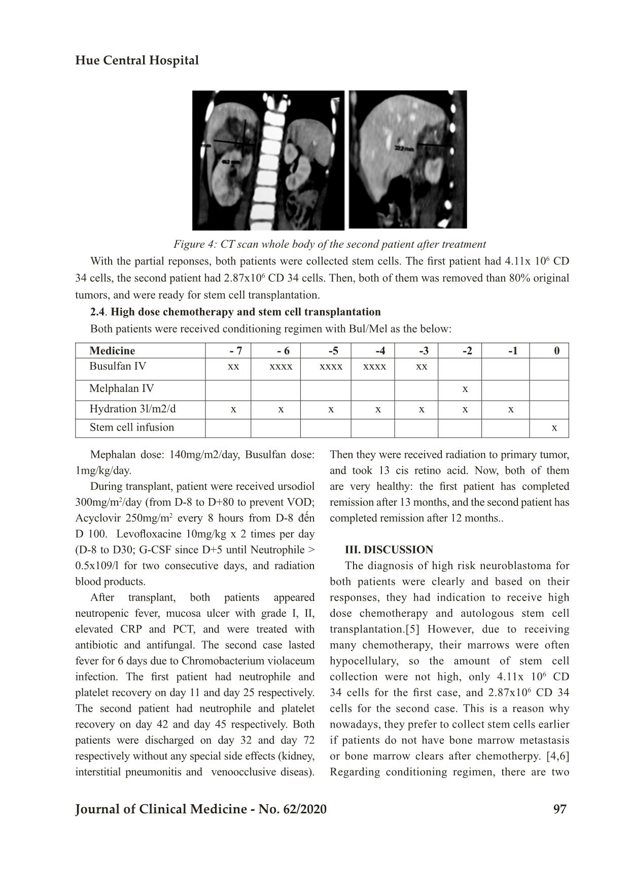
Figure 4: CT scan whole body of the second patient after treatment
With the partial reponses, both patients were collected stem cells. The first patient had 4.11x 10⁶ CD
34 cells, the second patient had 2.87x10⁶ CD 34 cells. Then, both of them was removed than 80% original
tumors, and were ready for stem cell transplantation.
2.4. High dose chemotherapy and stem cell transplantation
Both patients were received conditioning regimen with Bul/Mel as the below:
Medicine - 7 - 6 -5 -4 -3 -2 -1 0
Busulfan IV xx xxxx xxxx xxxx xx
Melphalan IV x
Hydration 3l/m2/d x x x x x x x
Stem cell infusion x
Mephalan dose: 140mg/m2/day, Busulfan dose:
1mg/kg/day.
During transplant, patient were received ursodiol
300mg/m2/day (from D-8 to D+80 to prevent VOD;
Acyclovir 250mg/m2 every 8 hours from D-8 đến
D 100. Levofloxacine 10mg/kg x 2 times per day
(D-8 to D30; G-CSF since D+5 until Neutrophile >
0.5x10⁹/l for two consecutive days, and radiation
blood products.
After transplant, both patients appeared
neutropenic fever, mucosa ulcer with grade I, II,
elevated CRP and PCT, and were treated with
antibiotic and antifungal. The second case lasted
fever for 6 days due to Chromobacterium violaceum
infection. The first patient had neutrophile and
platelet recovery on day 11 and day 25 respectively.
The second patient had neutrophile and platelet
recovery on day 42 and day 45 respectively. Both
patients were discharged on day 32 and day 72
respectively without any special side effects (kidney,
interstitial pneumonitis and venoocclusive diseas).
Then they were received radiation to primary tumor,
and took 13 cis retino acid. Now, both of them
are very healthy: the first patient has completed
remission after 13 months, and the second patient has
completed remission after 12 months..
III. DISCUSSION
The diagnosis of high risk neuroblastoma for
both patients were clearly and based on their
responses, they had indication to receive high
dose chemotherapy and autologous stem cell
transplantation.[5] However, due to receiving
many chemotherapy, their marrows were often
hypocellulary, so the amount of stem cell
collection were not high, only 4.11x 10⁶ CD
34 cells for the first case, and 2.87x10⁶ CD 34
cells for the second case. This is a reason why
nowadays, they prefer to collect stem cells earlier
if patients do not have bone marrow metastasis
or bone marrow clears after chemotherpy. [4,6]
Regarding conditioning regimen, there are two
Bệnh viện Trung ương Huế
98 Journal of Clinical Medicine - No. 62/2020
regimens: Bul/Mel (busulfan and melphalan) and
CEM (carboplatin, etoposide and melphalan).
[3,6,7] We chosed myeloablative therapy with
Bul/Mel followed by 13 cis retino-acid.[8,9] After
transplant, the first patient recoveried faster than
the second patient. This could be explained due to
the amount of CD 34 cell infusion. According to
Fish and Ali: the number of CD34+ cells infused
to a patient correlates with the likelihood of
successful engraftment. [4,10] The treatment was
well tolerated. Mucositis was the main toxicity,
with grade I and II. Both of them had developed
neutropenia fever and without any special
side effects, such as: renal effects, interstitial
pneumonitis and venooclusive disease.
IV. CONCLUSION
Management high risk neuroblastoma is
very challenge and need combined many drugs.
Autologous stem cell transplant plays an important
role in consolidation. This is the first two case
autologous stem cell transplant for high risk
neuroblastoma. Both of them tolerated well and had
no special side effects. Until now, they have one more
year survival since diagnosis. We continue to follow
up and implement the new therapy to more patients.
REFERENCES
1. Trahair TN, Vowels MR, Johnston K, Cohn
RJ, Russell SJ, Neville KA, et al. Long-
term outcomes in children with high-risk
neuroblastoma treated with autologous stem
cell transplantation. Bone Marrow Transplant
2007; 40: 741 - 6.
2. Lau SCD, Unni MNM, Teh KH, Aziz MA,
Muda Z, Thomas SA, et al. Autologous stem cell
transplantation following high-dose chemotherapy
in children with high-risk neuroblastoma:
Practicality in resource - limited countries. Pediatr
Blood Cancer 2020; 67: e28176.
3. Elzembely MM, Park JR, Riad KF, Sayed
HA, Pinto N, Carpenter PA, et al. Acute
Complications After High-Dose Chemotherapy
and Stem-Cell Rescue in Pediatric Patients With
High-Risk Neuroblastoma Treated in Countries
With Different Resources. J Glob Oncol 2018;
4: 1 - 12.
4. Fish JD, Grupp SA. Stem cell transplantation
for neuroblastoma. Bone Marrow Transplant
2008; 41: 159 - 65.
5. Matthay KK, Villablanca JG, Seeger RC, Stram
DO, Harris RE, Ramsay NK, et al. Treatment
of high - risk neuroblastoma with intensive
chemotherapy, radiotherapy, autologous bone
marrow transplantation, and 13 - cis - retinoic
acid. Children’s Cancer Group. N Engl J Med
1999; 341: 1165 - 73.
6. Soni S, Pai V, Gross TG, Ranalli M. Busulfan
and melphalan as consolidation therapy
with autologous peripheral blood stem cell
transplantation following Children’s Oncology
Group (COG) induction platform for high-
risk neuroblastoma: early results from a single
institution. Pediatr Transplant 2014; 18: 217 - 20.
7. Yalcin B, Kremer LC, Caron HN, van Dalen
EC. High-dose chemotherapy and autologous
haematopoietic stem cell rescue for children
with high - risk neuroblastoma. Cochrane
Database Syst Rev 2013: CD006301.
8. Parikh NS, Howard SC, Chantada G, Israels
T, Khattab M, Alcasabas P, et al. SIOP-PODC
adapted risk stratification and treatment
guidelines: Recommendations for neuroblastoma
in low- and middle-income settings. Pediatr
Blood Cancer 2015; 62: 1305 - 16.
9. Peinemann F, van Dalen EC, Enk H, Berthold
F. Retinoic acid postconsolidation therapy
for high-risk neuroblastoma patients treated
with autologous haematopoietic stem cell
transplantation. Cochrane Database Syst Rev
2017; 8: CD010685.
10. Ali MY, Oyama Y, Monreal J, Winter JN, Tallman
MS, Williams SF, et al. Ideal or actual body weight
to calculate CD34+ cell doses for autologous
hematopoietic stem cell transplantation? Bone
Marrow Transplant 2003; 31: 861 - 4
High dose chemotherapy and autologous...
File đính kèm:
 high_dose_chemotherapy_and_autologous_hematopoietic_stem_cel.pdf

