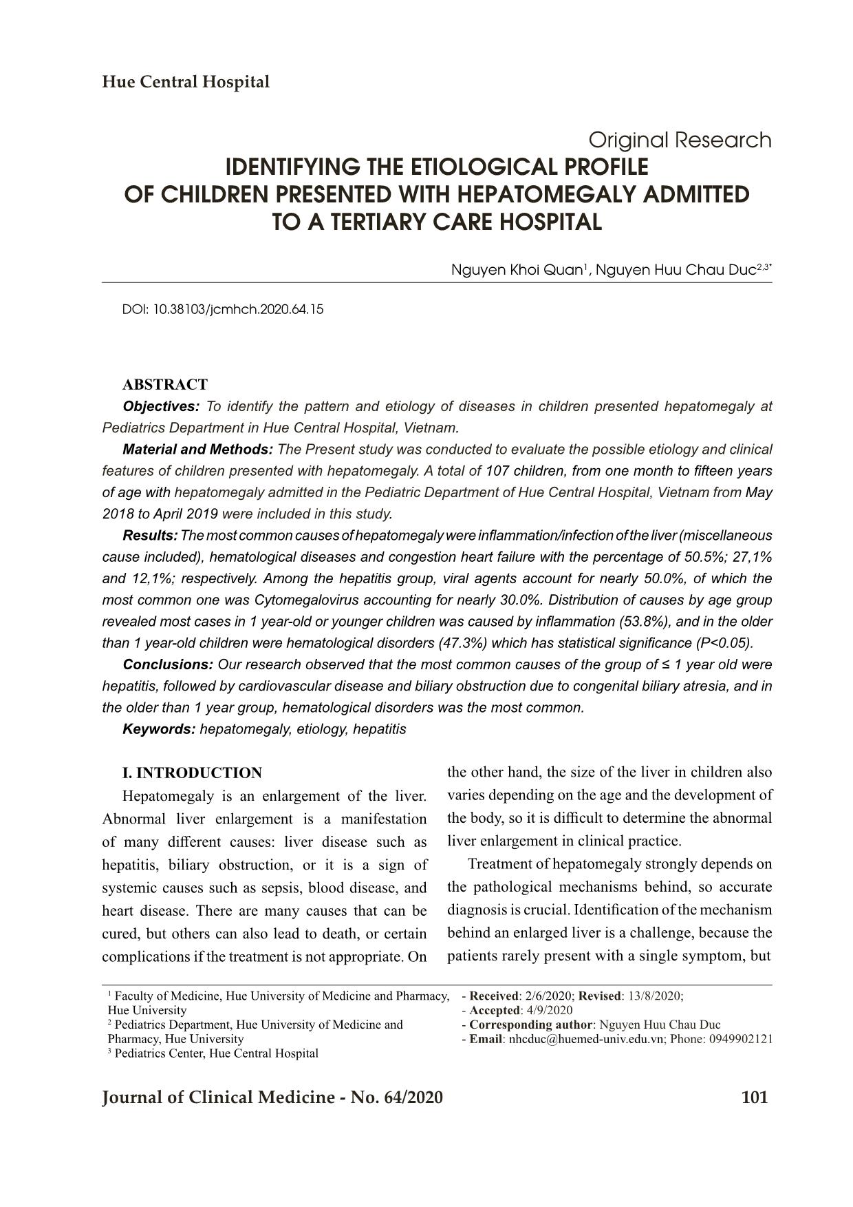
Dentifying the etiological profile of children presented with hepatomegaly admitted to a tertiary care hospital
Objectives: To identify the pattern and etiology of diseases in children presented hepatomegaly at
Pediatrics Department in Hue Central Hospital, Vietnam.
Material and Methods: The Present study was conducted to evaluate the possible etiology and clinical
features of children presented with hepatomegaly. A total of 107 children, from one month to fifteen years
of age with hepatomegaly admitted in the Pediatric Department of Hue Central Hospital, Vietnam from May
2018 to April 2019 were included in this study.
Results:The most common causes of hepatomegaly were inflammation/infection of the liver (miscellaneous
cause included), hematological diseases and congestion heart failure with the percentage of 50.5%; 27,1%
and 12,1%; respectively. Among the hepatitis group, viral agents account for nearly 50.0%, of which the
most common one was Cytomegalovirus accounting for nearly 30.0%. Distribution of causes by age group
revealed most cases in 1 year-old or younger children was caused by inflammation (53.8%), and in the older
than 1 year-old children were hematological disorders (47.3%) which has statistical significance (P<0.05).
Conclusions: Our research observed that the most common causes of the group of ≤ 1 year old were
hepatitis, followed by cardiovascular disease and biliary obstruction due to congenital biliary atresia, and in
the older than 1 year group, hematological disorders was the most common.
Trang 1
Trang 2
Trang 3
Trang 4
Trang 5
Trang 6
Trang 7
Trang 8
Trang 9
Tóm tắt nội dung tài liệu: Dentifying the etiological profile of children presented with hepatomegaly admitted to a tertiary care hospital
Hue Central Hospital
Journal of Clinical Medicine - No. 64/2020 101
Original Research
IDENTIFYING THE ETIOLOGICAL PROFILE
OF CHILDREN PRESENTED WITH HEPATOMEGALY ADMITTED
TO A TERTIARY CARE HOSPITAL
Nguyen Khoi Quan1, Nguyen Huu Chau Duc2,3*
DOI: 10.38103/jcmhch.2020.64.15
ABSTRACT
Objectives: To identify the pattern and etiology of diseases in children presented hepatomegaly at
Pediatrics Department in Hue Central Hospital, Vietnam.
Material and Methods: The Present study was conducted to evaluate the possible etiology and clinical
features of children presented with hepatomegaly. A total of 107 children, from one month to fifteen years
of age with hepatomegaly admitted in the Pediatric Department of Hue Central Hospital, Vietnam from May
2018 to April 2019 were included in this study.
Results: The most common causes of hepatomegaly were inflammation/infection of the liver (miscellaneous
cause included), hematological diseases and congestion heart failure with the percentage of 50.5%; 27,1%
and 12,1%; respectively. Among the hepatitis group, viral agents account for nearly 50.0%, of which the
most common one was Cytomegalovirus accounting for nearly 30.0%. Distribution of causes by age group
revealed most cases in 1 year-old or younger children was caused by inflammation (53.8%), and in the older
than 1 year-old children were hematological disorders (47.3%) which has statistical significance (P<0.05).
Conclusions: Our research observed that the most common causes of the group of ≤ 1 year old were
hepatitis, followed by cardiovascular disease and biliary obstruction due to congenital biliary atresia, and in
the older than 1 year group, hematological disorders was the most common.
Keywords: hepatomegaly, etiology, hepatitis
I. INTRODUCTION
Hepatomegaly is an enlargement of the liver.
Abnormal liver enlargement is a manifestation
of many different causes: liver disease such as
hepatitis, biliary obstruction, or it is a sign of
systemic causes such as sepsis, blood disease, and
heart disease. There are many causes that can be
cured, but others can also lead to death, or certain
complications if the treatment is not appropriate. On
the other hand, the size of the liver in children also
varies depending on the age and the development of
the body, so it is difficult to determine the abnormal
liver enlargement in clinical practice.
Treatment of hepatomegaly strongly depends on
the pathological mechanisms behind, so accurate
diagnosis is crucial. Identification of the mechanism
behind an enlarged liver is a challenge, because the
patients rarely present with a single symptom, but
1 Faculty of Medicine, Hue University of Medicine and Pharmacy,
Hue University
2 Pediatrics Department, Hue University of Medicine and
Pharmacy, Hue University
3 Pediatrics Center, Hue Central Hospital
- Received: 2/6/2020; Revised: 13/8/2020;
- Accepted: 4/9/2020
- Corresponding author: Nguyen Huu Chau Duc
- Email: nhcduc@huemed-univ.edu.vn; Phone: 0949902121
Bệnh viện Trung ương Huế
102 Journal of Clinical Medicine - No. 64/2020
often contemporary associated many symptoms. The
differences in many causes and their serious effects
on children also require early detection and prognosis
of the serious risk factors, in order to have an early
and appropriate treatment to prevent complications
and death. Currently, thanks to modern testing
techniques, most causes of hepatomegaly have been
discovered, and the disease has been prevented
and treated with efficiency. Unfortunately, some
laboratory tests are not available in many healthcare
facilities. A simple algorithm using a few common
parameters with easy-approach is needed in the
facilities and also for primary medical care.
All over the world as well as in Vietnam, studies
about the etiology of hepatomegaly is limited [1].
Most studies focus on one specific cause, separately
on each different study subject [2] [3]. In order to
contribute to the subject and improve the capacity
of diagnosis, we conducted the research with three
objectives: to reveal the etiology of hepatomegaly
in children; to describe the clinical and laboratory
characteristics of hepatomegaly children, and we
also present a clinical-approach algorithm to apply
into practice.
II. MATERIAL AND METHODS
A total 107 children from 1 moth to 15 years of
age admitted with hepatomegaly into the Pediatric
Department of Hue Central Hospital, Vietnam from
May 2018 to April 2019 included in the study.
Children with incomplete data were excluded from
this study. Those children presented with pushed
down liver due to various causes are excluded
by measuring liver span for that particular age.
Hepatomegaly was defined as when the liver edge
was palpated below the right costal margin at the
mid clavicular line for infants and older children
and the liver span should be more than the ex ... ent. Data
were collected from register book of the patient
by using data entry preform, which was prepared
by the researchers themselves. After collection
of relevant information the data were checked,
verified, edited manually for consistency, accuracy
and to reduce error. Statistical analysis was done
by statistical software SPSS for windows version
20. P values were calculated using Pearson chi-
square test. P < 0.05 was considered as significant
and P < 0.01 as highly significant.
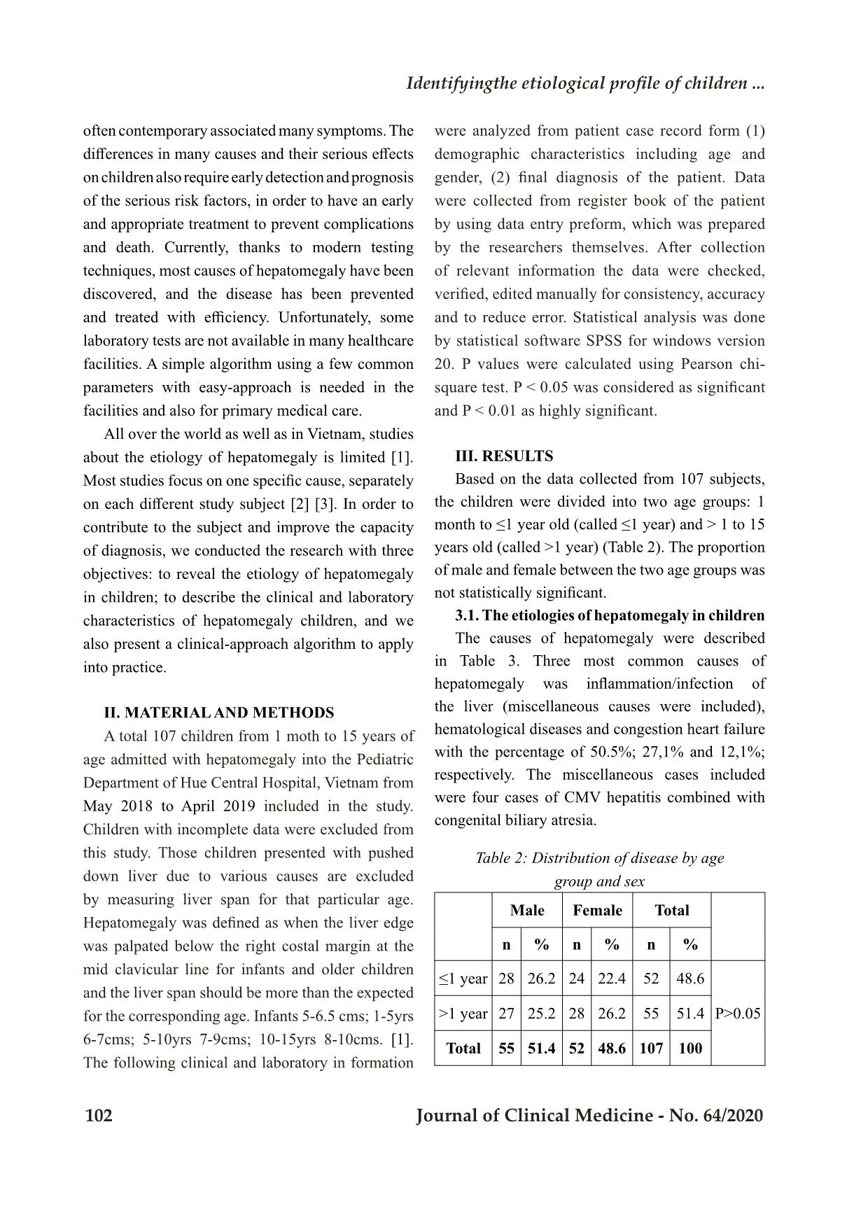
III. RESULTS
Based on the data collected from 107 subjects,
the children were divided into two age groups: 1
month to ≤1 year old (called ≤1 year) and > 1 to 15
years old (called >1 year) (Table 2). The proportion
of male and female between the two age groups was
not statistically significant.
3.1. The etiologies of hepatomegaly in children
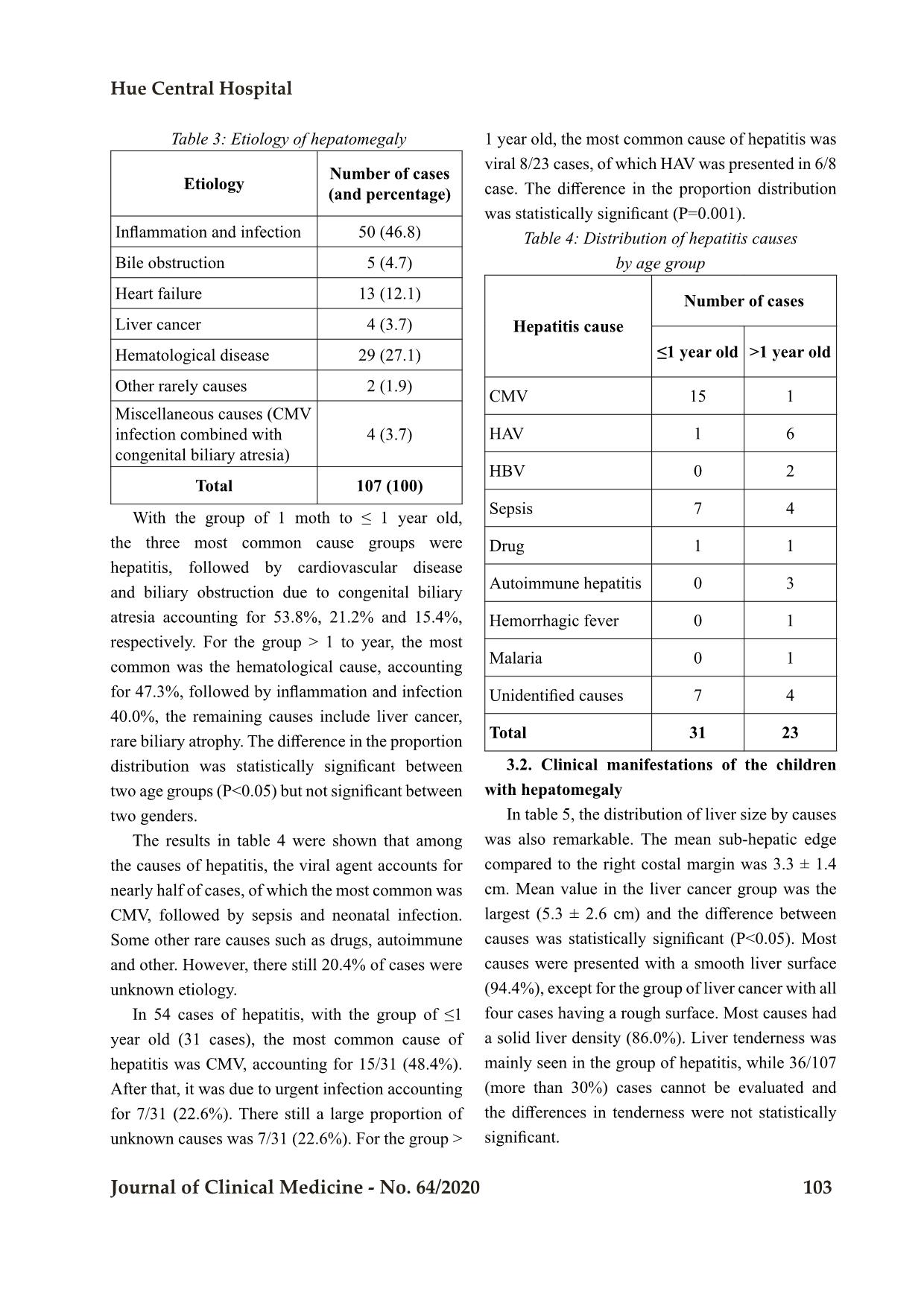
The causes of hepatomegaly were described
in Table 3. Three most common causes of
hepatomegaly was inflammation/infection of
the liver (miscellaneous causes were included),
hematological diseases and congestion heart failure
with the percentage of 50.5%; 27,1% and 12,1%;
respectively. The miscellaneous cases included
were four cases of CMV hepatitis combined with
congenital biliary atresia.
Table 2: Distribution of disease by age
group and sex
Male Female Total
n % n % n %
≤1 year 28 26.2 24 22.4 52 48.6
P>0.05>1 year 27 25.2 28 26.2 55 51.4
Total 55 51.4 52 48.6 107 100
Identifyingthe etiological profile of children ...
Hue Central Hospital
Journal of Clinical Medicine - No. 64/2020 103
 Table 3: Etiology of hepatomegaly
Etiology Number of cases (and percentage)
Inflammation and infection 50 (46.8)
Bile obstruction 5 (4.7)
Heart failure 13 (12.1)
Liver cancer 4 (3.7)
Hematological disease 29 (27.1)
Other rarely causes 2 (1.9)
Miscellaneous causes (CMV
infection combined with
congenital biliary atresia)
4 (3.7)
Total 107 (100)
With the group of 1 moth to ≤ 1 year old,
the three most common cause groups were
hepatitis, followed by cardiovascular disease
and biliary obstruction due to congenital biliary
atresia accounting for 53.8%, 21.2% and 15.4%,
respectively. For the group > 1 to year, the most
common was the hematological cause, accounting
for 47.3%, followed by inflammation and infection
40.0%, the remaining causes include liver cancer,
rare biliary atrophy. The difference in the proportion
distribution was statistically significant between
two age groups (P<0.05) but not significant between
two genders.
The results in table 4 were shown that among
the causes of hepatitis, the viral agent accounts for
nearly half of cases, of which the most common was
CMV, followed by sepsis and neonatal infection.
Some other rare causes such as drugs, autoimmune
and other. However, there still 20.4% of cases were
unknown etiology.
In 54 cases of hepatitis, with the group of ≤1
year old (31 cases), the most common cause of
hepatitis was CMV, accounting for 15/31 (48.4%).
After that, it was due to urgent infection accounting
for 7/31 (22.6%). There still a large proportion of
unknown causes was 7/31 (22.6%). For the group >
1 year old, the most common cause of hepatitis was
viral 8/23 cases, of which HAV was presented in 6/8
case. The difference in the proportion distribution
was statistically significant (P=0.001).
Table 4: Distribution of hepatitis causes
by age group
Hepatitis cause
Number of cases
≤1 year old >1 year old
CMV 15 1
HAV 1 6
HBV 0 2
Sepsis 7 4
Drug 1 1
Autoimmune hepatitis 0 3
Hemorrhagic fever 0 1
Malaria 0 1
Unidentified causes 7 4
Total 31 23
3.2. Clinical manifestations of the children
with hepatomegaly
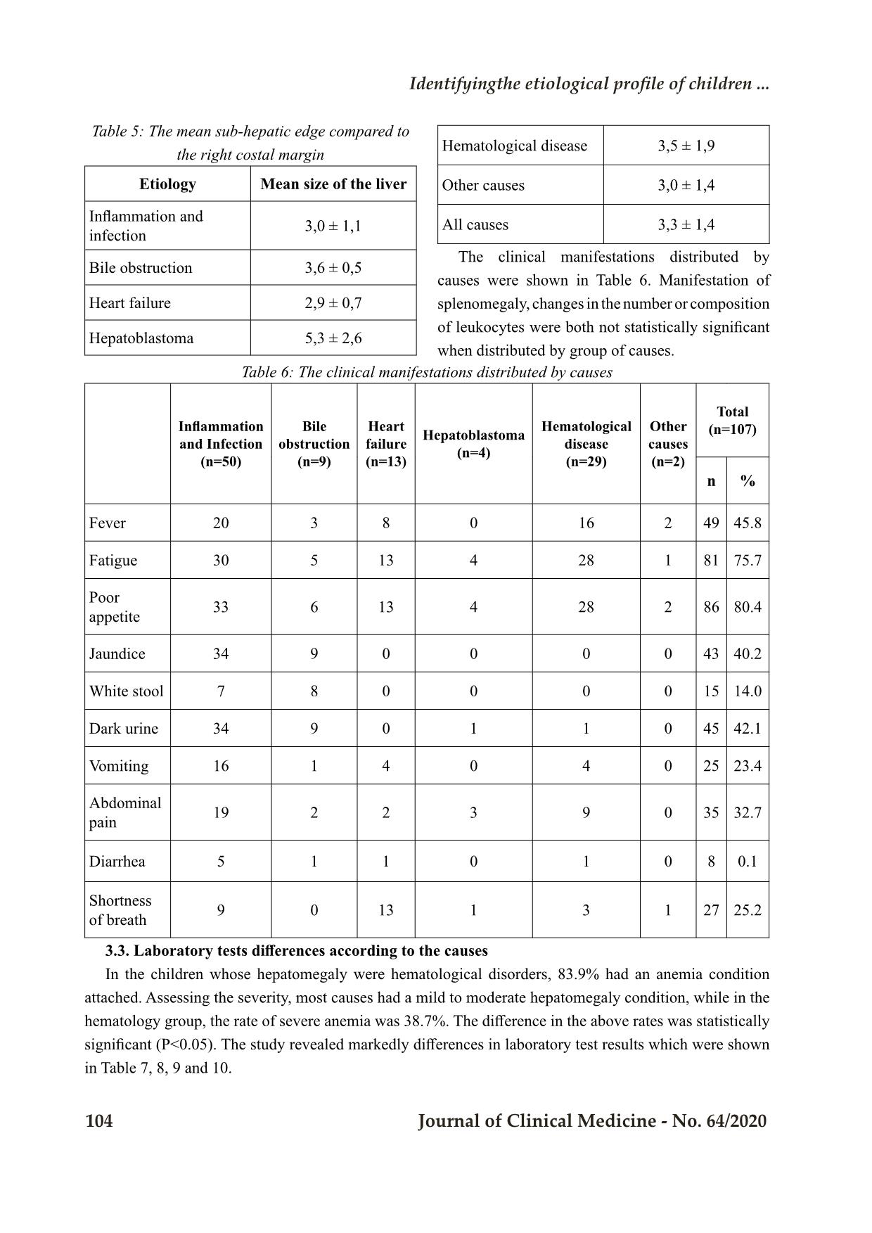
In table 5, the distribution of liver size by causes
was also remarkable. The mean sub-hepatic edge
compared to the right costal margin was 3.3 ± 1.4
cm. Mean value in the liver cancer group was the
largest (5.3 ± 2.6 cm) and the difference between
causes was statistically significant (P<0.05). Most
causes were presented with a smooth liver surface
(94.4%), except for the group of liver cancer with all
four cases having a rough surface. Most causes had
a solid liver density (86.0%). Liver tenderness was
mainly seen in the group of hepatitis, while 36/107
(more than 30%) cases cannot be evaluated and
the differences in tenderness were not statistically
significant.
Bệnh viện Trung ương Huế
104 Journal of Clinical Medicine - No. 64/2020
Table 5: The mean sub-hepatic edge compared to
the right costal margin
Etiology Mean size of the liver
Inflammation and
infection
3,0 ± 1,1
Bile obstruction 3,6 ± 0,5
Heart failure 2,9 ± 0,7
Hepatoblastoma 5,3 ± 2,6
Hematological disease 3,5 ± 1,9
Other causes 3,0 ± 1,4
All causes 3,3 ± 1,4
The clinical manifestations distributed by
causes were shown in Table 6. Manifestation of
splenomegaly, changes in the number or composition
of leukocytes were both not statistically significant
when distributed by group of causes.
Table 6: The clinical manifestations distributed by causes
Inflammation
and Infection
(n=50)
Bile
obstruction
(n=9)
Heart
failure
(n=13)
Hepatoblastoma
(n=4)
Hematological
disease
(n=29)
Other
causes
(n=2)
Total
(n=107)
n %
Fever 20 3 8 0 16 2 49 45.8
Fatigue 30 5 13 4 28 1 81 75.7
Poor
appetite 33 6 13 4 28 2 86 80.4
Jaundice 34 9 0 0 0 0 43 40.2
White stool 7 8 0 0 0 0 15 14.0
Dark urine 34 9 0 1 1 0 45 42.1
Vomiting 16 1 4 0 4 0 25 23.4
Abdominal
pain 19 2 2 3 9 0 35 32.7
Diarrhea 5 1 1 0 1 0 8 0.1
Shortness
of breath 9 0 13 1 3 1 27 25.2
3.3. Laboratory tests differences according to the causes
In the children whose hepatomegaly were hematological disorders, 83.9% had an anemia condition
attached. Assessing the severity, most causes had a mild to moderate hepatomegaly condition, while in the
hematology group, the rate of severe anemia was 38.7%. The difference in the above rates was statistically
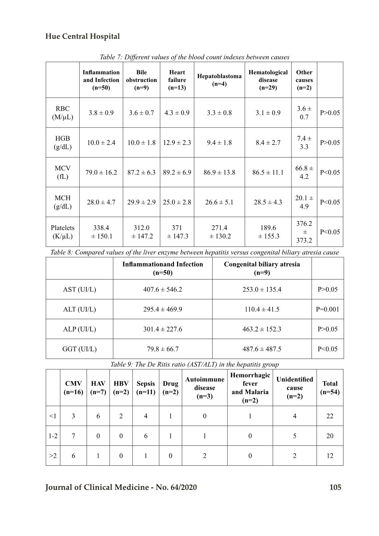
significant (P<0.05). The study revealed markedly differences in laboratory test results which were shown
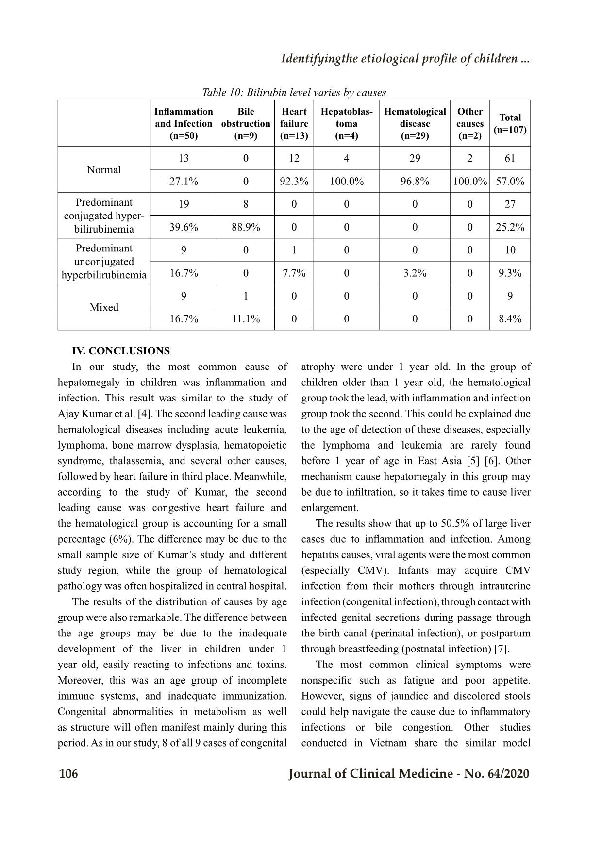
in Table 7, 8, 9 and 10.
Identifyingthe etiological profile of children ...
Hue Central Hospital
Journal of Clinical Medicine - No. 64/2020 105
Table 7: Different values of the blood count indexes between causes
Inflammation
and Infection
(n=50)
Bile
obstruction
(n=9)
Heart
failure
(n=13)
Hepatoblastoma
(n=4)
Hematological
disease
(n=29)
Other
causes
(n=2)
RBC
(M/µL)
3.8 ± 0.9 3.6 ± 0.7 4.3 ± 0.9 3.3 ± 0.8 3.1 ± 0.9
3.6 ±
0.7 P>0.05
HGB
(g/dL)
10.0 ± 2.4 10.0 ± 1.8 12.9 ± 2.3 9.4 ± 1.8 8.4 ± 2.7
7.4 ±
3.3 P>0.05
MCV
(fL)
79.0 ± 16.2 87.2 ± 6.3 89.2 ± 6.9 86.9 ± 13.8 86.5 ± 11.1
66.8 ±
4.2
P<0.05
MCH
(g/dL)
28.0 ± 4.7 29.9 ± 2.9 25.0 ± 2.8 26.6 ± 5.1 28.5 ± 4.3
20.1 ±
4.9
P<0.05
Platelets
(K/µL)
338.4
 ± 150.1
312.0
 ± 147.2
371
 ± 147.3
271.4
 ± 130.2
189.6
 ± 155.3
376.2
 ±
373.2
P<0.05
Table 8: Compared values of the liver enzyme between hepatitis versus congenital biliary atresia cause
Inflammationand Infection
(n=50)
Congenital biliary atresia
(n=9)
AST (UI/L) 407.6 ± 546.2 253.0 ± 135.4 P>0.05
ALT (UI/L) 295.4 ± 469.9 110.4 ± 41.5 P=0.001
ALP (UI/L) 301.4 ± 227.6 463.2 ± 152.3 P>0.05
GGT (UI/L) 79.8 ± 66.7 487.6 ± 487.5 P<0.05
Table 9: The De Ritis ratio (AST/ALT) in the hepatitis group
CMV
(n=16)
HAV
(n=7)
HBV
(n=2)
Sepsis
(n=11)
Drug
(n=2)
Autoimmune
disease
(n=3)
Hemorrhagic
fever
and Malaria
(n=2)
Unidentified
cause
(n=2)
Total
(n=54)
<1 3 6 2 4 1 0 1 4 22
1-2 7 0 0 6 1 1 0 5 20
>2 6 1 0 1 0 2 0 2 12
Bệnh viện Trung ương Huế
106 Journal of Clinical Medicine - No. 64/2020
Table 10: Bilirubin level varies by causes
Inflammation
and Infection
(n=50)
Bile
obstruction
(n=9)
Heart
failure
(n=13)
Hepatoblas-
toma
(n=4)
Hematological
disease
(n=29)
Other
causes
(n=2)
Total
(n=107)
Normal
13 0 12 4 29 2 61
27.1% 0 92.3% 100.0% 96.8% 100.0% 57.0%
Predominant
conjugated hyper-
bilirubinemia
19 8 0 0 0 0 27
39.6% 88.9% 0 0 0 0 25.2%
Predominant
unconjugated
hyperbilirubinemia
9 0 1 0 0 0 10
16.7% 0 7.7% 0 3.2% 0 9.3%
Mixed
9 1 0 0 0 0 9
16.7% 11.1% 0 0 0 0 8.4%
IV. CONCLUSIONS
In our study, the most common cause of
hepatomegaly in children was inflammation and
infection. This result was similar to the study of
Ajay Kumar et al. [4]. The second leading cause was
hematological diseases including acute leukemia,
lymphoma, bone marrow dysplasia, hematopoietic
syndrome, thalassemia, and several other causes,
followed by heart failure in third place. Meanwhile,
according to the study of Kumar, the second
leading cause was congestive heart failure and
the hematological group is accounting for a small
percentage (6%). The difference may be due to the
small sample size of Kumar’s study and different
study region, while the group of hematological
pathology was often hospitalized in central hospital.
The results of the distribution of causes by age
group were also remarkable. The difference between
the age groups may be due to the inadequate
development of the liver in children under 1
year old, easily reacting to infections and toxins.
Moreover, this was an age group of incomplete
immune systems, and inadequate immunization.
Congenital abnormalities in metabolism as well
as structure will often manifest mainly during this
period. As in our study, 8 of all 9 cases of congenital
atrophy were under 1 year old. In the group of
children older than 1 year old, the hematological
group took the lead, with inflammation and infection
group took the second. This could be explained due
to the age of detection of these diseases, especially
the lymphoma and leukemia are rarely found
before 1 year of age in East Asia [5] [6]. Other
mechanism cause hepatomegaly in this group may
be due to infiltration, so it takes time to cause liver
enlargement.
The results show that up to 50.5% of large liver
cases due to inflammation and infection. Among
hepatitis causes, viral agents were the most common
(especially CMV). Infants may acquire CMV
infection from their mothers through intrauterine
infection (congenital infection), through contact with
infected genital secretions during passage through
the birth canal (perinatal infection), or postpartum
through breastfeeding (postnatal infection) [7].
The most common clinical symptoms were
nonspecific such as fatigue and poor appetite.
However, signs of jaundice and discolored stools
could help navigate the cause due to inflammatory
infections or bile congestion. Other studies
conducted in Vietnam share the similar model
Identifyingthe etiological profile of children ...
Hue Central Hospital
Journal of Clinical Medicine - No. 64/2020 107
[8] [9]. Dyspnea were also valuable to suggest
heart failure as an underlying condition. We had
also evaluated accompanying splenomegaly, but
differences were not significant.
Changes in complete blood count were most
common in the hematological group, characterized
by a decrease in both erythrocytes and thrombocytes,
with an inconsistent sign of leukopenia. The MCV
alterations between the groups were remarkable.
These signs were consistent with pathogenesis of
liver malfunction. Due to the heterogeneousness
of tests, this study focused on differences between
the inflammation, infection and biliary obstruction
group. In the hepatitis group, increasing AST and
ALT were dominant than ALP and GGT; while
in the bile obstructive condition, in contrast,
ALP and GGT were markedly increased. This
difference was explained based on the origin of
these enzymes. AST, ALT in hepatitis children
reflects the destruction of hepatocytes. ALT mainly
originated from cytoplasm while AST was highly
localized in the mitochondria (80%) and a little in
cytoplasm (20%) therefore increasing AST suggests
deep and progressive damage. Beside, ALP was
synthesized by hepatocytes and bile duct epithelial
cells and GGT was found with high concentrations
in the cylindrical epithelial cells of the bile duct,
it reveals that ALP and GGT were very sensitive
tests to detect biliary obstruction. Bilirubin level
was normal in most causes. Bilirubin increase was
seen in all cases with biliary obstruction and also
in a large number of hepatitis. The obstructive
group had direct bilirubin dominance, while in the
hepatitis group the difference between the causes
was not really clear.
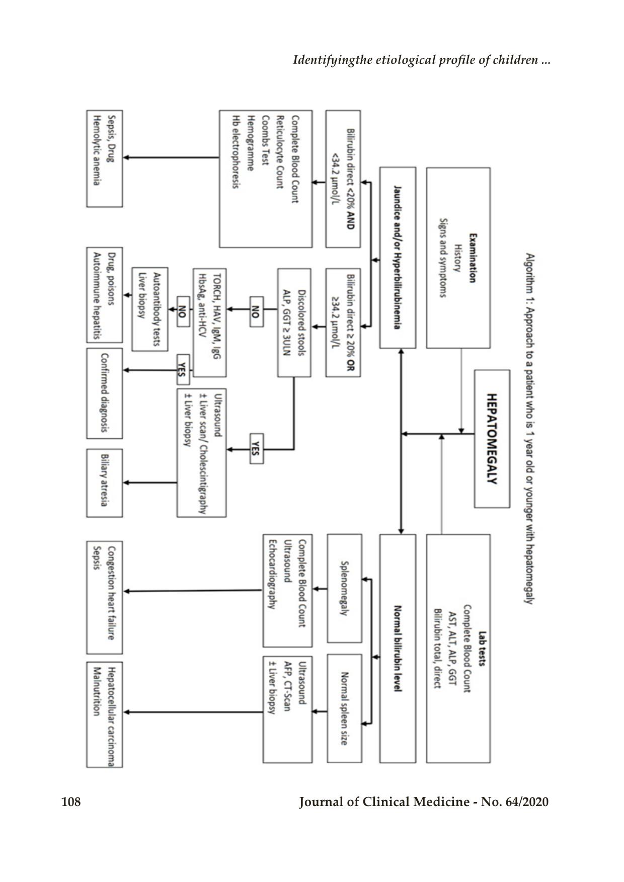
Our research strengthens the current
understanding of the etiology, clinical and
laboratory manifestations of hepatomegaly. Based
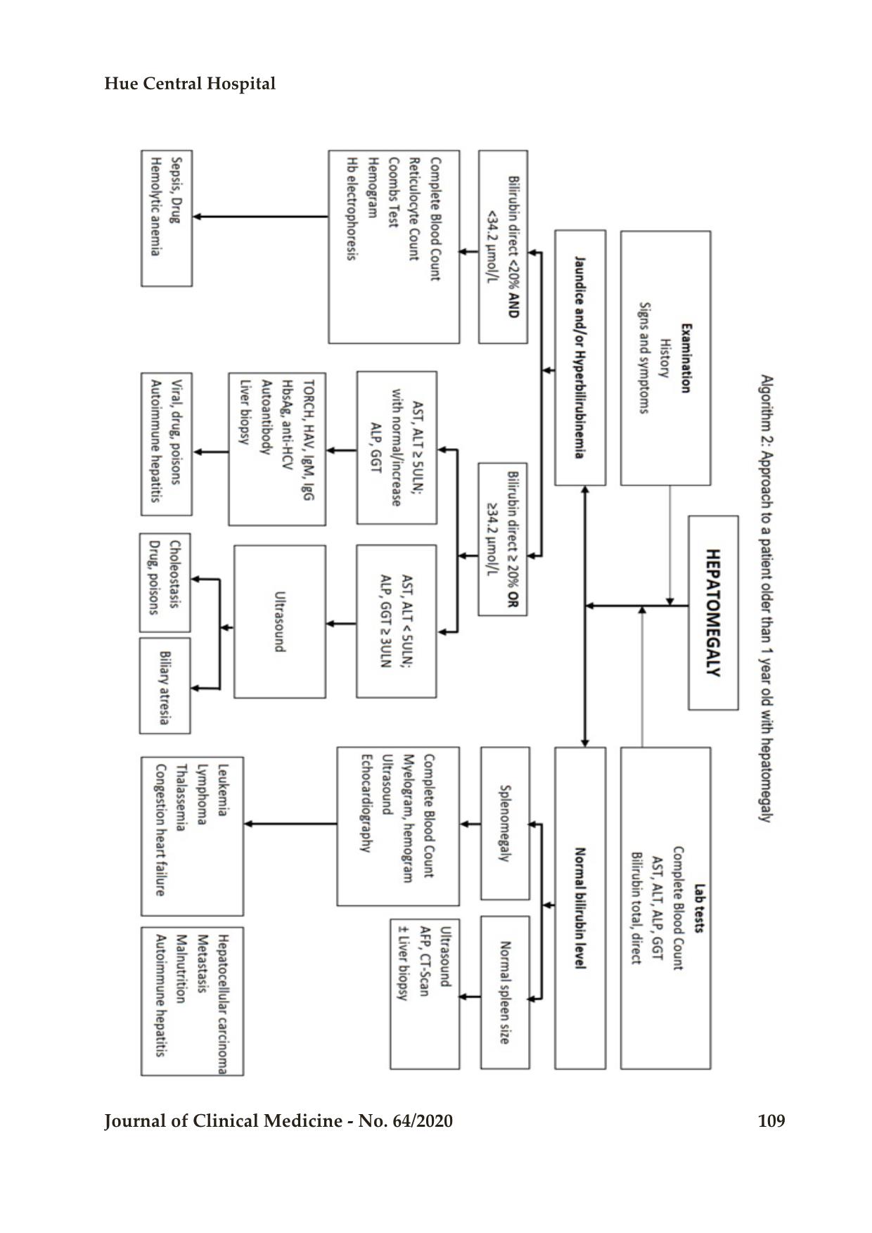
on these insights, we also propose two schemes for
approaching the diagnosis of hepatomegaly by two
age groups below (algorithm 1, 2).
Conflict of interests
All the authors have no conflict of interests to
declare.
REFERENCES
1. P. Song, ĐĐ. Đức, B. Hiền và cộng sự, “Đánh
giá bước đầu căn nguyên viêm gan virus ở Việt
Nam. Nghiên cứu lâm sàng căn nguyên học
viêm gan virus (1992-1996),” Đề tài KY 01-09,
04 1996.
2. CH. Hầu, “Nghiên cứu tình hình nhiễm viêm
gan siêu vi A, B và E trong cộng đồng tại huyện
Tân Châu tỉnh An Giang,” in Hội nghị khoa học
chuyên đề Vi sinh - Dịch tễ - Miễn dịch, Thành
phố Hồ Chí Minh, 1996.
3. NV. Nguyên, “Nghiên cứu lâm sàng, cận lâm
sàng và nguyên nhân của viêm gan cấp ở trẻ em
tại Bệnh viện Nhi Trung Ương,” Luận văn tốt
nghiệp thạc sỹ, Đại học Y Hà Nội, 2015.
4. A. Kumar, “Clinico pathological study of
hepatomegaly,” International Journal of
Medical and Health Research, vol. 2, no. 1, pp.
60-2, 2016.
5. BL Asselin, DG Tubergen, J Hochberg, “Cancer
and Benign Tumors,” in Nelson Textbook of
Pediatrics, 20th edition, New York, USA,
Elsevier, 2016, pp. 2416-80.
6. K. Nakata, Y. Ito, W. Magadi et al, “Childhood
cancer incidence and survival in Japan and
England: A population‐based study (1993‐2010),”
Cancer Science, vol. 109, no. 2, 2017.
7. MY Hasosah, SY Kutbi, AW Al-Amri et al,
“Perinatal cytomegalovirus hepatitis in Saudi
infants: a case series,” Saudi J Gastroenterol,
vol. 18, pp. 208-13, 2012.
8. [8] PN. An và cộng sự, “Đặc điểm lâm sàng,
cận lâm sàng, dịch tễ học và diễn biến của viêm
gan virus cấp ở trẻ em trong 5 năm 1991-1995
tại Viện Nhi Trung Ương,” Nhi khoa 7(1), pp.
12-9, 1998.
9. VTH. Yến, “Tìm hiểu căn nguyên, đặc điểm lâm
sàng và biến đổi một số chỉ số huyết học, hoá
sinh trong viêm gan virus cấp trẻ em tại Bệnh
viện Nhi Trung Ương,” Luận văn tốt nghiệp
thạc sĩ, Đại học Y Hà Nội, 2003.
Bệnh viện Trung ương Huế
108 Journal of Clinical Medicine - No. 64/2020
Identifyingthe etiological profile of children ...
Hue Central Hospital
Journal of Clinical Medicine - No. 64/2020 109
File đính kèm:
 dentifying_the_etiological_profile_of_children_presented_wit.pdf

