Characteristic of inflammatory myofibrinoblastic tumor: Retrospective analysis of 4 cases in children hospital number 2
Introduction: Inflammatory myofibroblastic tumor (IMT) is a rare mesenchymal tumor that involves
various organs. Surgery is the mainstay of therapy for this tumor. Approximately half of all IMT cases have
anaplastic lymphoma kinase (ALK) rearrangements; therefore, the ALK inhibitor crizotinib is suggested as a
promising treatment for unresectable cases with ALK rearrangements. In cases withoutALK rearrangement,
chemotherapy is an alternative treatment for unresectable tumors.
Cases report: We report 4 cases of IMT treated in Children Hospital Number 2 since 2018. There was
one case with mesenteric tumor, one with perineal tumor detected at birth, one with inferior mediastinal
tumor and one with intestinal tumor. We evaluated ALK expression by immunohistochemistry and ALK
rearrangement by fluorescence in situ hybridization (FISH) in 3 cases and all negative. Treatments included
tumor resection or biopsy and chemotherapy with methotrexate (30mg/m2) day 1 and vincristine (1.5mg/
m2) day 1 and 7 in a 3 week cycle together with NSAID or steroid for inflammatory control. There was one
case with complete response to surgery and chemotherapy; the other 3 cases ended in death due to tumor
recurrence (3 cases) and metastasis (1 case).
Conclusions: IMT has a diverse clinical presentation, appears in many different locations, and the
clinical course can progress quickly with high rate of recurrence after incomplete surgical resection. Surgery
is the optimal approach, but in those without complete resection and in those with tumor progression,
tyrosine kinase inhibition should be considered if there is ALK rearrangement. In the absence of ALK
rearrangement, additional tyrosine kinase rearrangements and other potentially efficacious chemotherapy
regimens need to be studied for these patients.
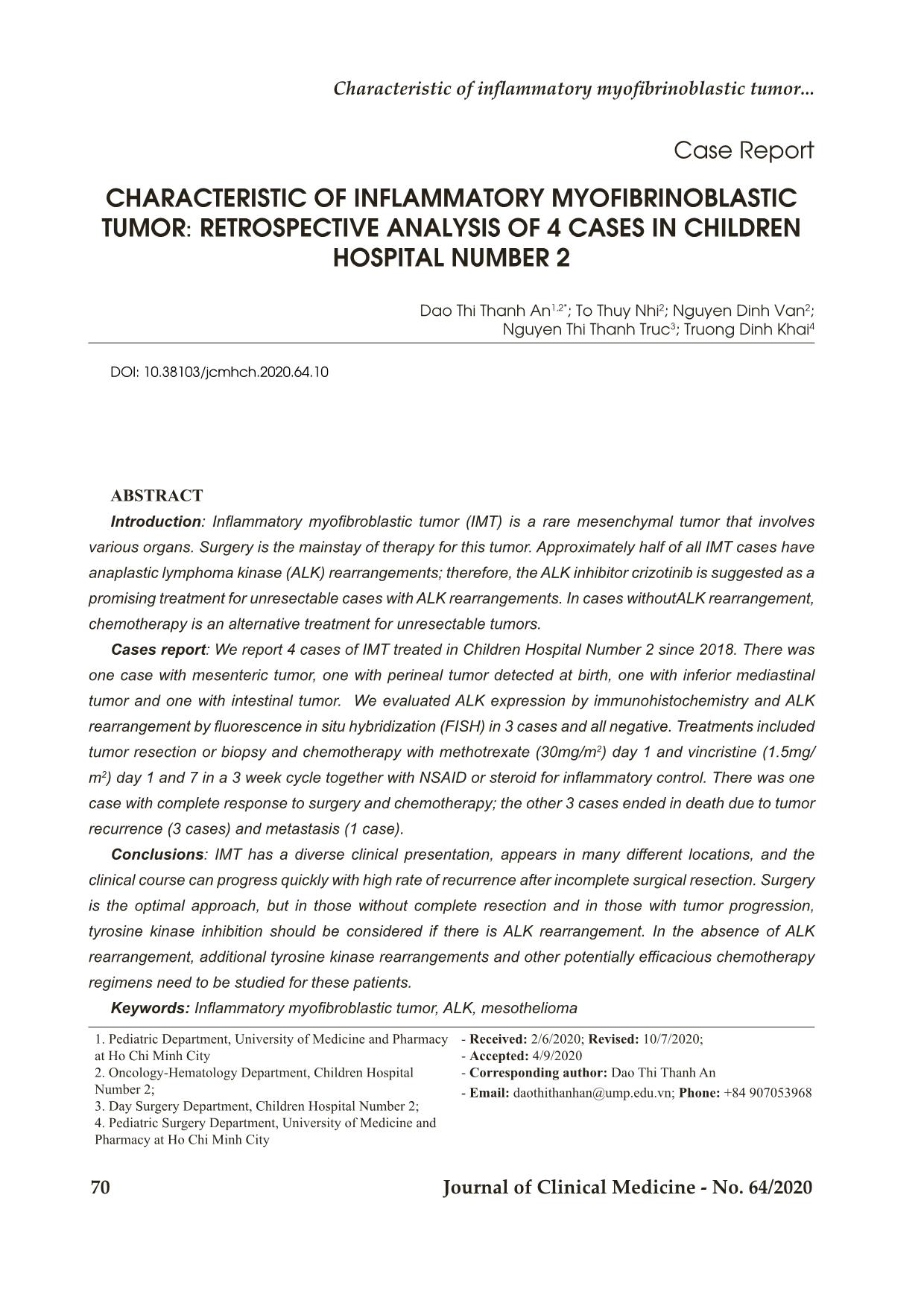
Trang 1
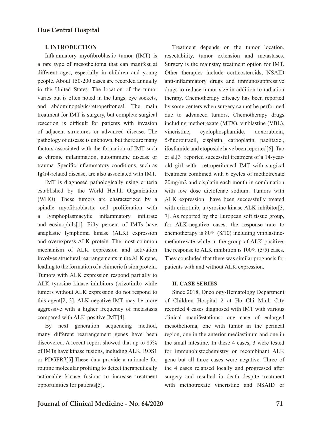
Trang 2
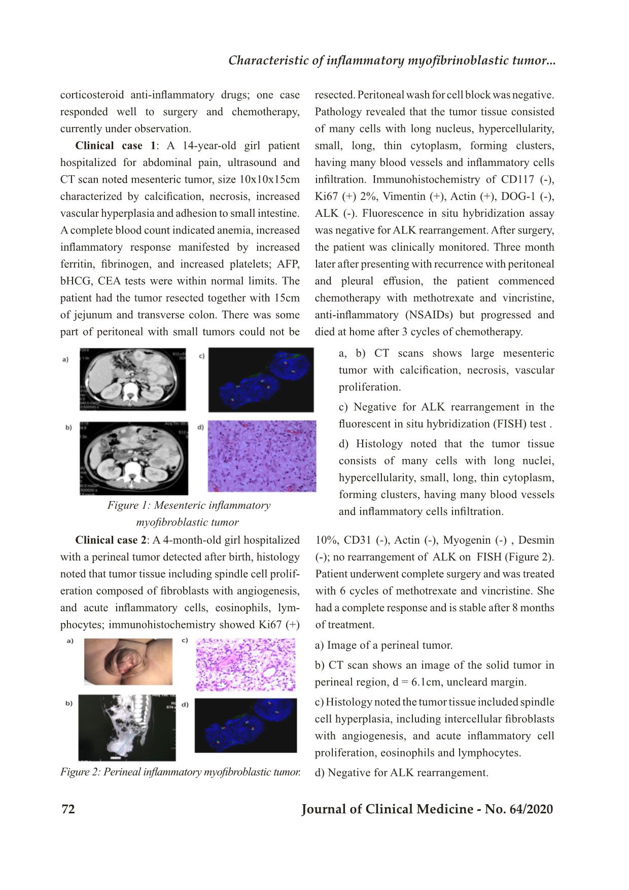
Trang 3
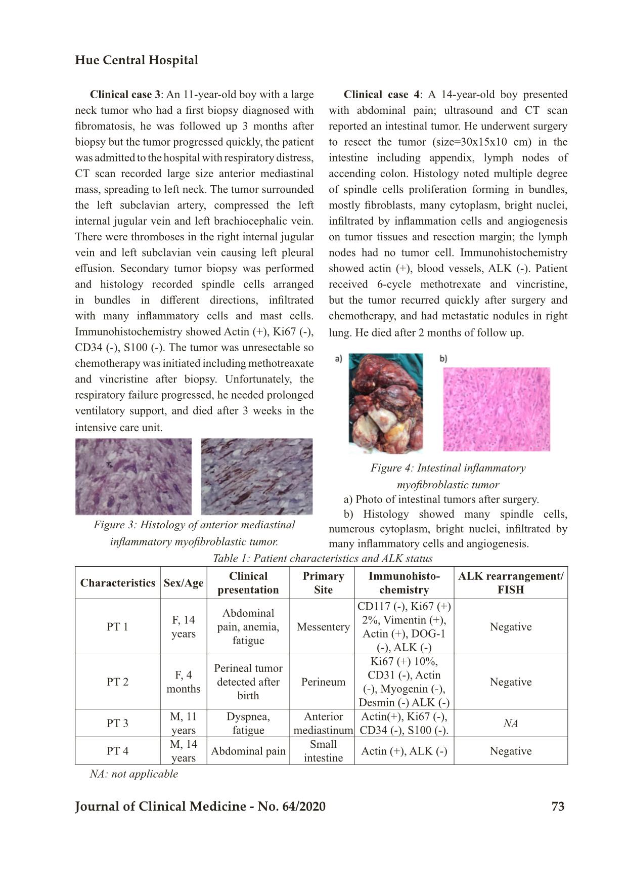
Trang 4
Trang 5
Trang 6
Tóm tắt nội dung tài liệu: Characteristic of inflammatory myofibrinoblastic tumor: Retrospective analysis of 4 cases in children hospital number 2
Bệnh viện Trung ương Huế
70 Journal of Clinical Medicine - No. 64/2020
Case Report
CHARACTERISTIC OF INFLAMMATORY MYOFIBRINOBLASTIC
TUMOR: RETROSPECTIVE ANALYSIS OF 4 CASES IN CHILDREN
HOSPITAL NUMBER 2
Dao Thi Thanh An1,2*; To Thuy Nhi2; Nguyen Dinh Van2;
Nguyen Thi Thanh Truc3; Truong Dinh Khai4
DOI: 10.38103/jcmhch.2020.64.10
ABSTRACT
Introduction: Inflammatory myofibroblastic tumor (IMT) is a rare mesenchymal tumor that involves
various organs. Surgery is the mainstay of therapy for this tumor. Approximately half of all IMT cases have
anaplastic lymphoma kinase (ALK) rearrangements; therefore, the ALK inhibitor crizotinib is suggested as a
promising treatment for unresectable cases with ALK rearrangements. In cases withoutALK rearrangement,
chemotherapy is an alternative treatment for unresectable tumors.
Cases report: We report 4 cases of IMT treated in Children Hospital Number 2 since 2018. There was
one case with mesenteric tumor, one with perineal tumor detected at birth, one with inferior mediastinal
tumor and one with intestinal tumor. We evaluated ALK expression by immunohistochemistry and ALK
rearrangement by fluorescence in situ hybridization (FISH) in 3 cases and all negative. Treatments included
tumor resection or biopsy and chemotherapy with methotrexate (30mg/m2) day 1 and vincristine (1.5mg/
m2) day 1 and 7 in a 3 week cycle together with NSAID or steroid for inflammatory control. There was one
case with complete response to surgery and chemotherapy; the other 3 cases ended in death due to tumor
recurrence (3 cases) and metastasis (1 case).
Conclusions: IMT has a diverse clinical presentation, appears in many different locations, and the
clinical course can progress quickly with high rate of recurrence after incomplete surgical resection. Surgery
is the optimal approach, but in those without complete resection and in those with tumor progression,
tyrosine kinase inhibition should be considered if there is ALK rearrangement. In the absence of ALK
rearrangement, additional tyrosine kinase rearrangements and other potentially efficacious chemotherapy
regimens need to be studied for these patients.
Keywords: Inflammatory myofibroblastic tumor, ALK, mesothelioma
1. Pediatric Department, University of Medicine and Pharmacy
at Ho Chi Minh City
2. Oncology-Hematology Department, Children Hospital
Number 2;
3. Day Surgery Department, Children Hospital Number 2;
4. Pediatric Surgery Department, University of Medicine and
Pharmacy at Ho Chi Minh City
- Received: 2/6/2020; Revised: 10/7/2020;
- Accepted: 4/9/2020
- Corresponding author: Dao Thi Thanh An
- Email: daothithanhan@ump.edu.vn; Phone: +84 907053968
Characteristic of inflammatory myofibri oblastic tumor...
Hue Central Hospital
Journal of Clinical Medicine - No. 64/2020 71
I. INTRODUCTION
Inflammatory myofibroblastic tumor (IMT) is
a rare type of mesothelioma that can manifest at
different ages, especially in children and young
people. About 150-200 cases are recorded annually
in the United States. The location of the tumor
varies but is often noted in the lungs, eye sockets,
and abdominopelvic/retroperitoneal. The main
treatment for IMT is surgery, but complete surgical
resection is difficult for patients with invasion
of adjacent structures or advanced disease. The
pathology of disease is unknown, but there are many
factors associated with the formation of IMT such
as chronic inflammation, autoimmune disease or
trauma. Specific inflammatory conditions, such as
IgG4-related disease, are also associated with IMT.
IMT is diagnosed pathologically using criteria
established by the World Health Organization
(WHO). These tumors are characterized by a
spindle myofibroblastic cell proliferation with
a lymphoplasmacytic inflammatory infiltrate
and eosinophils[1]. Fifty percent of IMTs have
anaplastic lymphoma kinase (ALK) expression
and overexpress ALK protein. The most common
mechanism of ALK expression and activation
involves structural rearrangements in the ALK gene,
leading to the formation of a chimeric fusion protein.
Tumors with ALK expression respond partially to
ALK tyrosine kinase inhibitors (crizotinib) while
tumors without ALK expression do not respond to
this agent[2, 3]. ALK-negative IMT may be more
aggressive with a higher frequency of metastasis
compared with ALK-positive IMT[4].
By next generation sequencing method,
many different rearrangement genes have been
discovered. A recent report showed that up to 85%
of IMTs have kinase fusions, including ALK, ROS1
or PDGFRβ[5].These data provide a rationale for
routine molecular profiling to detect therapeutically
actionable kinase fusions to increase treatment
opportunities for patients[5].
Treatment depends on the tumor location,
resectability, tumor extension and metastases.
Surgery is the mainstay treatment option for IMT.
Other therapies include corticosteroids, NSAID
anti-inflammatory drugs and immunosuppressive
drugs to reduce tumor size in addition to radiation
therapy. Chemotherapy efficacy has been reported
by some centers when surgery cannot be performed
due to advanced tumors. Chemotherapy drugs
including methotrexate (MTX), vinblastine (VBL),
vincristine, cyclophosphamide, doxorubicin,
5-fluorouracil, cisplatin, carboplatin, paclitaxel,
ifosfamide and etoposide have been reported[6]. Tao
et al.[3] reported successful treatment of a 14-year-
old girl with retroperitoneal IMT with surgical
treatment combined with 6 cycles of methotrexate
20mg/m2 and cisplatin each month in combination
with low dose diclofenac sodium. Tumors with
ALK expression have been successfully treated
with crizotinib, a tyrosine kinase ALK inhibitor[3,
7]. As reported by the European soft tissue group,
for ALK-negative cases, the response rate to
chemotherapy is 80% (8/10) including vinblastine-
methotrexate while in the group of ALK positive,
the response to ALK inhibition is 100% (5/5) cases.
They concluded that there was similar prognosis for
patients with and without ALK expression.
II. CASE SERIES
Since 2018, Oncology-Hematology Department
of Children Hospital 2 at Ho Chi Minh City
recorded 4 cases diagnosed with IMT with various
clinical manifestations: one case of enlarged
mesothelioma, one with tumor in the perineal
region, one in the anterior mediastinum and one in
the small intestine. In these 4 cases, 3 were tested
for immunohistochemistry or recombinant ALK
gene but all three cases were negative. Three of
the 4 cases relapsed locally and progressed after
surgery and resulted in death despite treatment
with methotrexate vincristine and NSAID or
1. Pediatric Department, University of Medicine and Pharmacy
at Ho Chi Minh City
2. Oncology-Hematology Department, Children Hospital
Number 2;
3. Day Surgery Department, Children Hospital Number 2;
4. Pediatric Surgery Department, University of Medicine and
Pharmacy at Ho Chi Minh City
- Received: 2/6/2020; Revised: 10/7/2020;
- Accepted: 4/9/2020
- Corresponding author: Dao Thi Thanh An
- Email: daothithanhan@ump.edu.vn; Phone: +84 907053968
Bệnh viện Trung ương Huế
72 Journal of Clinical Medicine - No. 64/2020
corticosteroid anti-inflammatory drugs; one case
responded well to surgery and chemotherapy,
currently under observation.
Clinical case 1: A 14-year-old girl patient
hospitalized for abdominal pain, ultrasound and
CT scan noted mesenteric tumor, size 10x10x15cm
characterized by calcification, necrosis, increased
vascular hyperplasia and adhesion to small intestine.
A complete blood count indicated anemia, increased
inflammatory response manifested by increased
ferritin, fibrinogen, and increased platelets; AFP,
bHCG, CEA tests were within normal limits. The
patient had the tumor resected together with 15cm
of jejunum and transverse colon. There was some
part of peritoneal with small tumors could not be
resected. Peritoneal wash for cell block was negative.
Pathology revealed that the tumor tissue consisted
of many cells with long nucleus, hypercellularity,
small, long, thin cytoplasm, forming clusters,
having many blood vessels and inflammatory cells
infiltration. Immunohistochemistry of CD117 (-),
Ki67 (+) 2%, Vimentin (+), Actin (+), DOG-1 (-),
ALK (-). Fluorescence in situ hybridization assay
was negative for ALK rearrangement. After surgery,
the patient was clinically monitored. Three month
later after presenting with recurrence with peritoneal
and pleural effusion, the patient commenced
chemotherapy with methotrexate and vincristine,
anti-inflammatory (NSAIDs) but progressed and
died at home after 3 cycles of chemotherapy.
a, b) CT scans shows large mesenteric
tumor with calcification, necrosis, vascular
proliferation.
c) Negative for ALK rearrangement in the
fluorescent in situ hybridization (FISH) test .
d) Histology noted that the tumor tissue
consists of many cells with long nuclei,
hypercellularity, small, long, thin cytoplasm,
forming clusters, having many blood vessels
and inflammatory cells infiltration.
a) Image of a perineal tumor.
b) CT scan shows an image of the solid tumor in
perineal region, d = 6.1cm, uncleard margin.
c) Histology noted the tumor tissue included spindle
cell hyperplasia, including intercellular fibroblasts
with angiogenesis, and acute inflammatory cell
proliferation, eosinophils and lymphocytes.
d) Negative for ALK rearrangement.
 Figure 1: Mesenteric inflammatory
myofibroblastic tumor
Clinical case 2: A 4-month-old girl hospitalized
with a perineal tumor detected after birth, histology
noted that tumor tissue including spindle cell prolif-
eration composed of fibroblasts with angiogenesis,
and acute inflammatory cells, eosinophils, lym-
phocytes; immunohistochemistry showed Ki67 (+)
10%, CD31 (-), Actin (-), Myogenin (-) , Desmin
(-); no rearrangement of ALK on FISH (Figure 2).
Patient underwent complete surgery and was treated
with 6 cycles of methotrexate and vincristine. She
had a complete response and is stable after 8 months
of treatment.
Figure 2: Perineal inflammatory myofibroblastic tumor.
Characteristic of inflammatory myofibri oblastic tumor...
Hue Central Hospital
Journal of Clinical Medicine - No. 64/2020 73
Clinical case 3: An 11-year-old boy with a large
neck tumor who had a first biopsy diagnosed with
fibromatosis, he was followed up 3 months after
biopsy but the tumor progressed quickly, the patient
was admitted to the hospital with respiratory distress,
CT scan recorded large size anterior mediastinal
mass, spreading to left neck. The tumor surrounded
the left subclavian artery, compressed the left
internal jugular vein and left brachiocephalic vein.
There were thromboses in the right internal jugular
vein and left subclavian vein causing left pleural
effusion. Secondary tumor biopsy was performed
and histology recorded spindle cells arranged
in bundles in different directions, infiltrated
with many inflammatory cells and mast cells.
Immunohistochemistry showed Actin (+), Ki67 (-),
CD34 (-), S100 (-). The tumor was unresectable so
chemotherapy was initiated including methotreaxate
and vincristine after biopsy. Unfortunately, the
respiratory failure progressed, he needed prolonged
ventilatory support, and died after 3 weeks in the
intensive care unit.
Clinical case 4: A 14-year-old boy presented
with abdominal pain; ultrasound and CT scan
reported an intestinal tumor. He underwent surgery
to resect the tumor (size=30x15x10 cm) in the
intestine including appendix, lymph nodes of
accending colon. Histology noted multiple degree
of spindle cells proliferation forming in bundles,
mostly fibroblasts, many cytoplasm, bright nuclei,
infiltrated by inflammation cells and angiogenesis
on tumor tissues and resection margin; the lymph
nodes had no tumor cell. Immunohistochemistry
showed actin (+), blood vessels, ALK (-). Patient
received 6-cycle methotrexate and vincristine,
but the tumor recurred quickly after surgery and
chemotherapy, and had metastatic nodules in right
lung. He died after 2 months of follow up.
Figure 3: Histology of anterior mediastinal
inflammatory myofibroblastic tumor.
a) Photo of intestinal tumors after surgery.
b) Histology showed many spindle cells,
numerous cytoplasm, bright nuclei, infiltrated by
many inflammatory cells and angiogenesis.
Figure 4: Intestinal inflammatory
myofibroblastic tumor
 Table 1: Patient characteristics and ALK status
Characteristics Sex/Age Clinical presentation
Primary
Site
Immunohisto-
chemistry
ALK rearrangement/
FISH
PT 1 F, 14 years
Abdominal
pain, anemia,
fatigue
Messentery
CD117 (-), Ki67 (+)
2%, Vimentin (+),
Actin (+), DOG-1
(-), ALK (-)
Negative
PT 2 F, 4 months
Perineal tumor
detected after
birth
Perineum
Ki67 (+) 10%,
CD31 (-), Actin
(-), Myogenin (-),
Desmin (-) ALK (-)
Negative
PT 3 M, 11 years
Dyspnea,
fatigue
Anterior
mediastinum
Actin(+), Ki67 (-),
CD34 (-), S100 (-).
NA
PT 4 M, 14 years Abdominal pain
Small
intestine Actin (+), ALK (-) Negative
NA: not applicable
Bệnh viện Trung ương Huế
74 Journal of Clinical Medicine - No. 64/2020
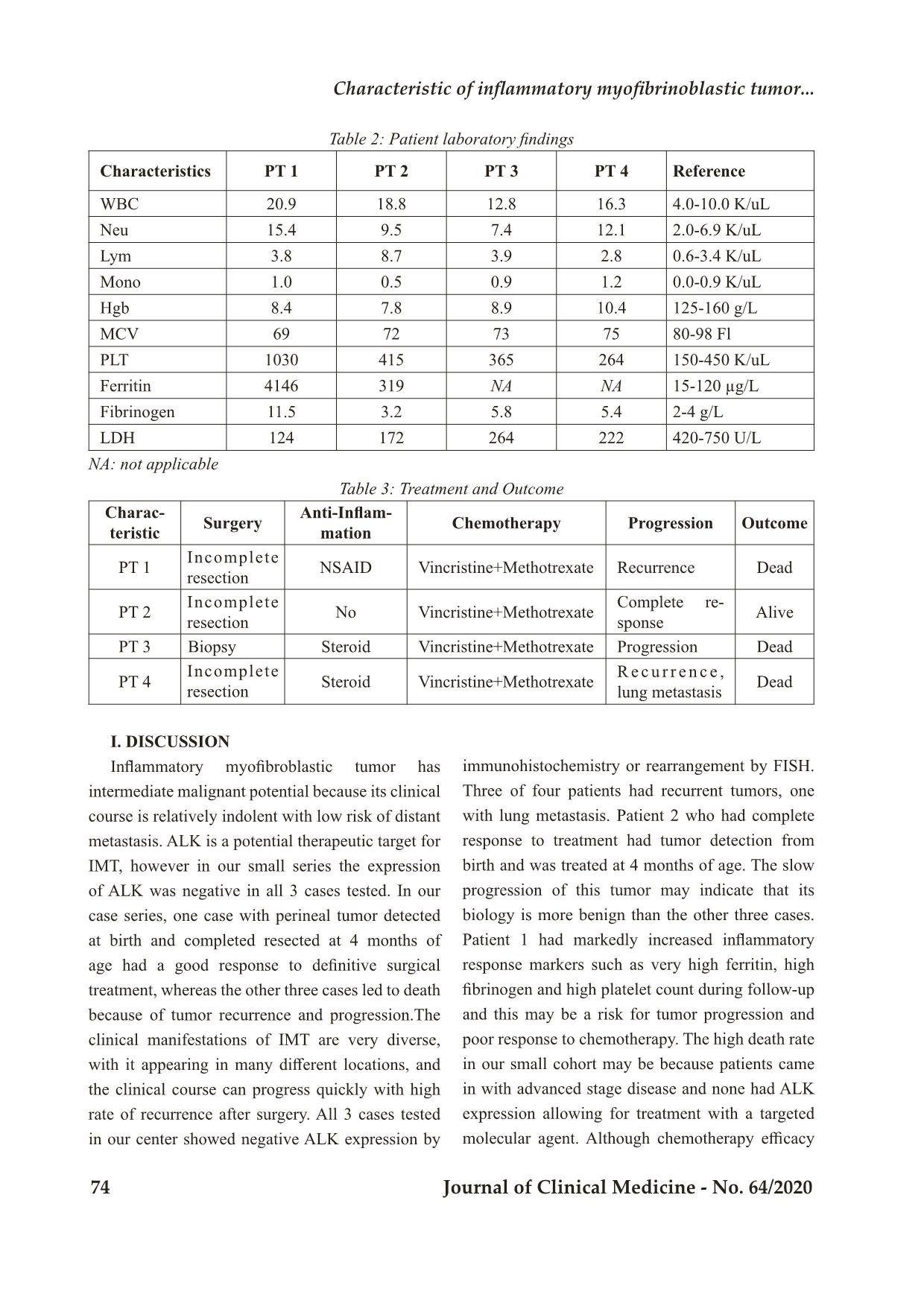
Table 2: Patient laboratory findings
Characteristics PT 1 PT 2 PT 3 PT 4 Reference
WBC 20.9 18.8 12.8 16.3 4.0-10.0 K/uL
Neu 15.4 9.5 7.4 12.1 2.0-6.9 K/uL
Lym 3.8 8.7 3.9 2.8 0.6-3.4 K/uL
Mono 1.0 0.5 0.9 1.2 0.0-0.9 K/uL
Hgb 8.4 7.8 8.9 10.4 125-160 g/L
MCV 69 72 73 75 80-98 Fl
PLT 1030 415 365 264 150-450 K/uL
Ferritin 4146 319 NA NA 15-120 µg/L
Fibrinogen 11.5 3.2 5.8 5.4 2-4 g/L
LDH 124 172 264 222 420-750 U/L
NA: not applicable
Table 3: Treatment and Outcome
Charac-
teristic Surgery
Anti-Inflam-
mation Chemotherapy Progression Outcome
PT 1 Incomplete resection NSAID Vincristine+Methotrexate Recurrence Dead
PT 2 Incomplete resection No Vincristine+Methotrexate
Complete re-
sponse Alive
PT 3 Biopsy Steroid Vincristine+Methotrexate Progression Dead
PT 4 Incomplete resection Steroid Vincristine+Methotrexate
R e c u r r e n c e ,
lung metastasis Dead
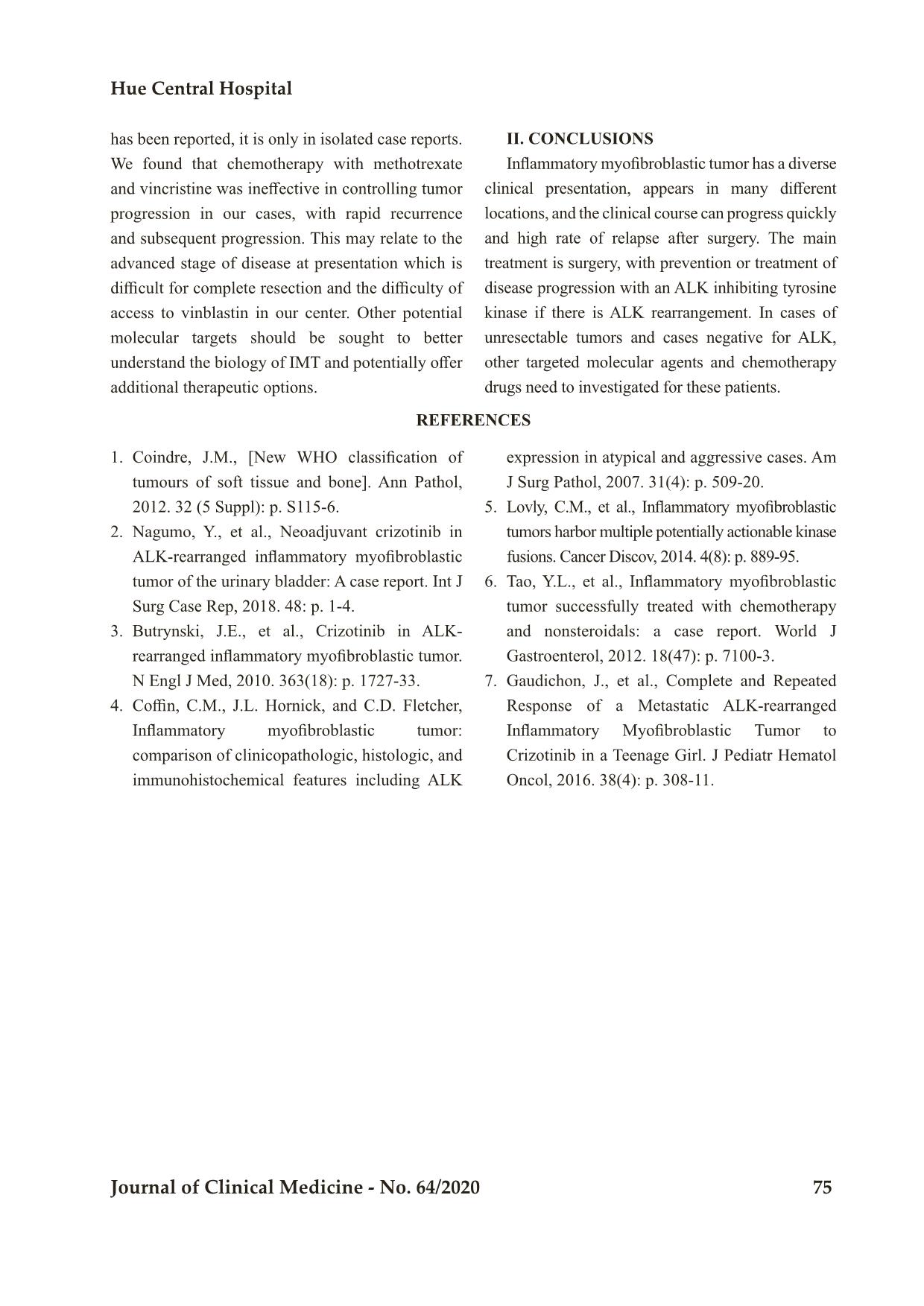
I. DISCUSSION
Inflammatory myofibroblastic tumor has
intermediate malignant potential because its clinical
course is relatively indolent with low risk of distant
metastasis. ALK is a potential therapeutic target for
IMT, however in our small series the expression
of ALK was negative in all 3 cases tested. In our
case series, one case with perineal tumor detected
at birth and completed resected at 4 months of
age had a good response to definitive surgical
treatment, whereas the other three cases led to death
because of tumor recurrence and progression.The
clinical manifestations of IMT are very diverse,
with it appearing in many different locations, and
the clinical course can progress quickly with high
rate of recurrence after surgery. All 3 cases tested
in our center showed negative ALK expression by
immunohistochemistry or rearrangement by FISH.
Three of four patients had recurrent tumors, one
with lung metastasis. Patient 2 who had complete
response to treatment had tumor detection from
birth and was treated at 4 months of age. The slow
progression of this tumor may indicate that its
biology is more benign than the other three cases.
Patient 1 had markedly increased inflammatory
response markers such as very high ferritin, high
fibrinogen and high platelet count during follow-up
and this may be a risk for tumor progression and
poor response to chemotherapy. The high death rate
in our small cohort may be because patients came
in with advanced stage disease and none had ALK
expression allowing for treatment with a targeted
molecular agent. Although chemotherapy efficacy
Characteristic of inflammatory myofibri oblastic tumor...
Hue Central Hospital
Journal of Clinical Medicine - No. 64/2020 75
has been reported, it is only in isolated case reports.
We found that chemotherapy with methotrexate
and vincristine was ineffective in controlling tumor
progression in our cases, with rapid recurrence
and subsequent progression. This may relate to the
advanced stage of disease at presentation which is
difficult for complete resection and the difficulty of
access to vinblastin in our center. Other potential
molecular targets should be sought to better
understand the biology of IMT and potentially offer
additional therapeutic options.
II. CONCLUSIONS
Inflammatory myofibroblastic tumor has a diverse
clinical presentation, appears in many different
locations, and the clinical course can progress quickly
and high rate of relapse after surgery. The main
treatment is surgery, with prevention or treatment of
disease progression with an ALK inhibiting tyrosine
kinase if there is ALK rearrangement. In cases of
unresectable tumors and cases negative for ALK,
other targeted molecular agents and chemotherapy
drugs need to investigated for these patients.
REFERENCES
1. Coindre, J.M., [New WHO classification of
tumours of soft tissue and bone]. Ann Pathol,
2012. 32 (5 Suppl): p. S115-6.
2. Nagumo, Y., et al., Neoadjuvant crizotinib in
ALK-rearranged inflammatory myofibroblastic
tumor of the urinary bladder: A case report. Int J
Surg Case Rep, 2018. 48: p. 1-4.
3. Butrynski, J.E., et al., Crizotinib in ALK-
rearranged inflammatory myofibroblastic tumor.
N Engl J Med, 2010. 363(18): p. 1727-33.
4. Coffin, C.M., J.L. Hornick, and C.D. Fletcher,
Inflammatory myofibroblastic tumor:
comparison of clinicopathologic, histologic, and
immunohistochemical features including ALK
expression in atypical and aggressive cases. Am
J Surg Pathol, 2007. 31(4): p. 509-20.
5. Lovly, C.M., et al., Inflammatory myofibroblastic
tumors harbor multiple potentially actionable kinase
fusions. Cancer Discov, 2014. 4(8): p. 889-95.
6. Tao, Y.L., et al., Inflammatory myofibroblastic
tumor successfully treated with chemotherapy
and nonsteroidals: a case report. World J
Gastroenterol, 2012. 18(47): p. 7100-3.
7. Gaudichon, J., et al., Complete and Repeated
Response of a Metastatic ALK-rearranged
Inflammatory Myofibroblastic Tumor to
Crizotinib in a Teenage Girl. J Pediatr Hematol
Oncol, 2016. 38(4): p. 308-11.
File đính kèm:
 characteristic_of_inflammatory_myofibrinoblastic_tumor_retro.pdf

