Castleman disease in children: Diagnosis and treatment
Background: We describe the experiences in diagnosis and results of treatment in pediatric Castleman
disease.
Method: Serial case reports.
Result: From 2016 to 2019, we had 7 cases of pediatric Castleman disease: 3 boys and 4 girl. The
median age at diagnosis was 147 months (121-173 months). Clinical manifestations were found in five
cases. They were all unicentric Castleman disease (6 abdominal mass, one left infraclavicular mass). All
patients were operated with postoperative period uneventful. The median time of postoperative follow up
was 22.7 months (11-53 months) with no signs of relapse.
Conclusions: Pediatric Castleman disease is a rare benign lymph node hyperplasia, it can be localised
or disseminated. Operation is the treatment of choice for localised Castleman disease.
Key words: Castleman disease, angiofollicular lymph node hyperplasia, giant lymph node hyperplasia,
lymph node hamartoma.
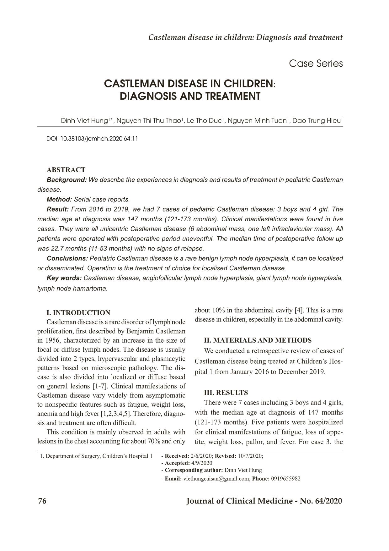
Trang 1
Trang 2
Trang 3
Trang 4
Trang 5
Trang 6
Tóm tắt nội dung tài liệu: Castleman disease in children: Diagnosis and treatment
Bệnh viện Trung ương Huế
76 Journal of Clinical Medicine - No. 64/2020
Case Series
CASTLEMAN DISEASE IN CHILDREN:
DIAGNOSIS AND TREATMENT
Dinh Viet Hung1*, Nguyen Thi Thu Thao1, Le Tho Duc1, Nguyen Minh Tuan1, Dao Trung Hieu1
DOI: 10.38103/jcmhch.2020.64.11
ABSTRACT
Background: We describe the experiences in diagnosis and results of treatment in pediatric Castleman
disease.
Method: Serial case reports.
Result: From 2016 to 2019, we had 7 cases of pediatric Castleman disease: 3 boys and 4 girl. The
median age at diagnosis was 147 months (121-173 months). Clinical manifestations were found in five
cases. They were all unicentric Castleman disease (6 abdominal mass, one left infraclavicular mass). All
patients were operated with postoperative period uneventful. The median time of postoperative follow up
was 22.7 months (11-53 months) with no signs of relapse.
Conclusions: Pediatric Castleman disease is a rare benign lymph node hyperplasia, it can be localised
or disseminated. Operation is the treatment of choice for localised Castleman disease.
Key words: Castleman disease, angiofollicular lymph node hyperplasia, giant lymph node hyperplasia,
lymph node hamartoma.
1. Department of Surgery, Children’s Hospital 1 - Received: 2/6/2020; Revised: 10/7/2020;
- Accepted: 4/9/2020
- Corresponding author: Dinh Viet Hung
- Email: viethungcaisan@gmail.com; Phone: 0919655982
I. INTRODUCTION
Castleman disease is a rare disorder of lymph node
proliferation, first described by Benjamin Castleman
in 1956, characterized by an increase in the size of
focal or diffuse lymph nodes. The disease is usually
divided into 2 types, hypervascular and plasmacytic
patterns based on microscopic pathology. The dis-
ease is also divided into localized or diffuse based
on general lesions [1-7]. Clinical manifestations of
Castleman disease vary widely from asymptomatic
to nonspecific features such as fatigue, weight loss,
anemia and high fever [1,2,3,4,5]. Therefore, diagno-
sis and treatment are often difficult.
This condition is mainly observed in adults with
lesions in the chest accounting for about 70% and only
about 10% in the abdominal cavity [4]. This is a rare
disease in children, especially in the abdominal cavity.
II. MATERIALS AND METHODS
We conducted a retrospective review of cases of
Castleman disease being treated at Children’s Hos-
pital 1 from January 2016 to December 2019.
III. RESULTS
There were 7 cases including 3 boys and 4 girls,
with the median age at diagnosis of 147 months
(121-173 months). Five patients were hospitalized
for clinical manifestations of fatigue, loss of appe-
tite, weight loss, pallor, and fever. For case 3, the
Castleman disease in children: Diagnosis and treatment
Hue Central Hospital
Journal of Clinical Medicine - No. 64/2020 77
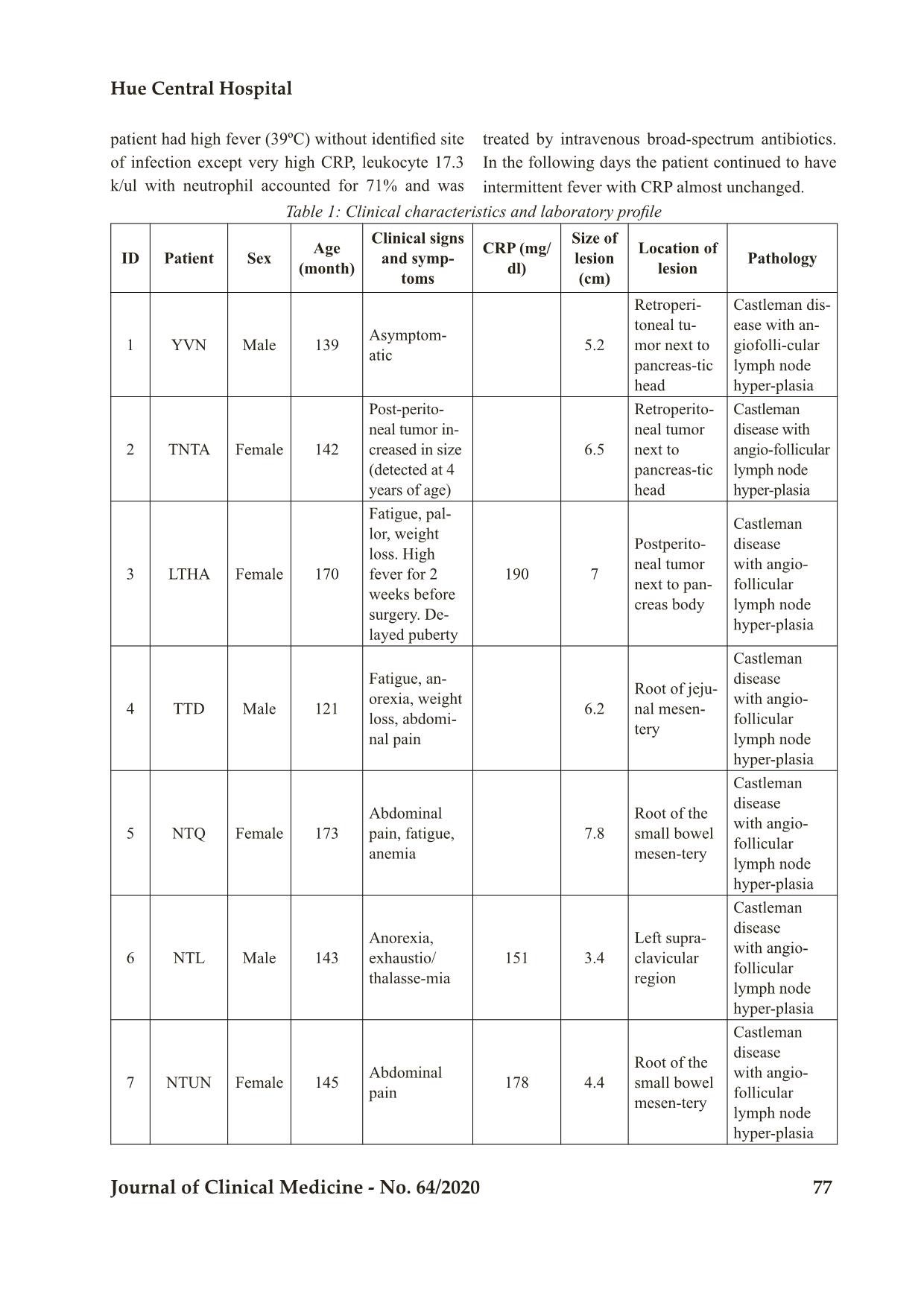
Table 1: Clinical characteristics and laboratory profile
ID Patient Sex Age (month)
Clinical signs
and symp-
toms
CRP (mg/
dl)
Size of
lesion
(cm)
Location of
lesion Pathology
1 YVN Male 139 Asymptom-atic 5.2
Retroperi-
toneal tu-
mor next to
pancreas-tic
head
Castleman dis-
ease with an-
giofolli-cular
lymph node
hyper-plasia
2 TNTA Female 142
Post-perito-
neal tumor in-
creased in size
(detected at 4
years of age)
6.5
Retroperito-
neal tumor
next to
pancreas-tic
head
Castleman
disease with
angio-follicular
lymph node
hyper-plasia
3 LTHA Female 170
Fatigue, pal-
lor, weight
loss. High
fever for 2
weeks before
surgery. De-
layed puberty
190 7
Postperito-
neal tumor
next to pan-
creas body
Castleman
disease
with angio-
follicular
lymph node
hyper-plasia
4 TTD Male 121
Fatigue, an-
orexia, weight
loss, abdomi-
nal pain
6.2
Root of jeju-
nal mesen-
tery
Castleman
disease
with angio-
follicular
lymph node
hyper-plasia
5 NTQ Female 173
Abdominal
pain, fatigue,
anemia
7.8
Root of the
small bowel
mesen-tery
Castleman
disease
with angio-
follicular
lymph node
hyper-plasia
6 NTL Male 143
Anorexia,
exhaustio/
thalasse-mia
151 3.4
Left supra-
clavicular
region
Castleman
disease
with angio-
follicular
lymph node
hyper-plasia
7 NTUN Female 145 Abdominal pain 178 4.4
Root of the
small bowel
mesen-tery
Castleman
disease
with angio-
follicular
lymph node
hyper-plasia
patient had high fever (39ºC) without identified site
of infection except very high CRP, leukocyte 17.3
k/ul with neutrophil accounted for 71% and was
treated by intravenous broad-spectrum antibiotics.
In the following days the patient continued to have
intermittent fever with CRP almost unchanged.
Bệnh viện Trung ương Huế
78 Journal of Clinical Medicine - No. 64/2020
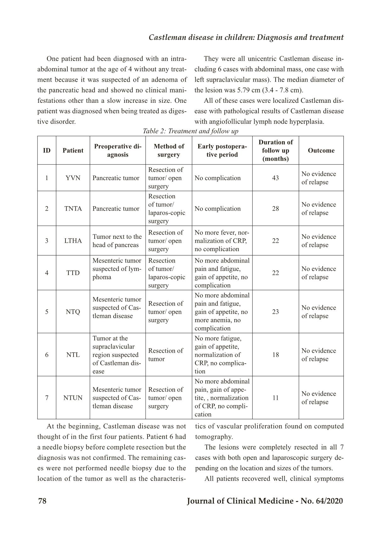
Table 2: Treatment and follow up
ID Patient Preoperative di-agnosis
Method of
surgery
Early postopera-
tive period
Duration of
follow up
(months)
Outcome
1 YVN Pancreatic tumor
Resection of
tumor/ open
surgery
No complication 43 No evidence of relapse
2 TNTA Pancreatic tumor
Resection
of tumor/
laparos-copic
surgery
No complication 28 No evidence of relapse
3 LTHA Tumor next to the head of pancreas
Resection of
tumor/ open
surgery
No more fever, nor-
malization of CRP,
no complication
22 No evidence of relapse
4 TTD
Mesenteric tumor
suspected of lym-
phoma
Resection
of tumor/
laparos-copic
surgery
No more abdominal
pain and fatigue,
gain of appetite, no
complication
22 No evidence of relapse
5 NTQ
Mesenteric tumor
suspected of Cas-
tleman disease
Resection of
tumor/ open
surgery
No more abdominal
pain and fatigue,
gain of appetite, no
more anemia, no
complication
23 No evidence of relapse
6 NTL
Tumor at the
supraclavicular
region suspected
of Castleman dis-
ease
Resection of
tumor
No more fatigue,
gain of appetite,
normalization of
CRP, no complica-
tion
18 No evidence of relapse
7 NTUN
Mesenteric tumor
suspected of Cas-
tleman disease
Resection of
tumor/ open
surgery
No more abdominal
pain, gain of appe-
tite, , normalization
of CRP, no compli-
cation
11 No evidence of relapse
One patient had been diagnosed with an intra-
abdominal tumor at the age of 4 without any treat-
ment because it was suspected of an adenoma of
the pancreatic head and showed no clinical mani-
festations other than a slow increase in size. One
patient was diagnosed when being treated as diges-
tive disorder.
They were all unicentric Castleman disease in-
cluding 6 cases with abdominal mass, one case with
left supraclavicular mass). The median diameter of
the lesion was 5.79 cm (3.4 - 7.8 cm).
All of these cases were localized Castleman dis-
ease with pathological results of Castleman disease
with angiofollicular lymph node hyperplasia.
At the beginning, Castleman disease was not
thought of in the first four patients. Patient 6 had
a needle biopsy before complete resection but the
diagnosis was not confirmed. The remaining cas-
es were not performed needle biopsy due to the
location of the tumor as well as the characteris-
tics of vascular proliferation found on computed
tomography.
The lesions were completely resected in all 7
cases with both open and laparoscopic surgery de-
pending on the location and sizes of the tumors.
All patients recovered well, clinical symptoms
Castleman disease in children: Diagnosis and treatment
Hue Central Hospital
Journal of Clinical Medicine - No. 64/2020 79
quickly disappeared right in the first few days after
surgery. Antibiotics for cases with fever were dis-
continued as soon as the results of pathological find-
ings were obtained and CRP levels decreased to 33
mg/dl on the third postoperative day.
These patients were discharged and monitored
based on clinical manifestations and periodic ul-
trasound examination with a median follow-up
time of 22.7 months (11-53 months). Results at
follow-up showed a physical recovery, gain of ap-
petite as well as the disappearance of symptoms
and no signs of relapse.
IV. DISCUSSION
Castleman disease is a benign proliferation of
lymphocytes and cytoplasm, also known by other
names: giant lymph node hyperplasia, lymph
node hamartoma, angiofollicular lymph node
hyperplasia [4].
Since the first case with lesions locating in the an-
terior mediastinum in adults was described by Cas-
tleman et al in 1954, an increasing number of cases
have been reported with various locations of the lym-
phatic system. Castleman disease appears to occur in
any location of the body’s lymphatic system, with the
most common site being the anterior mediastinum
accounting for about 70%, only less than 10% in the
abdominal cavity followed by the neck [2,4].
Six out of 7 cases in our report had intra-ab-
dominal lesions, perhaps a coincidence to help us
learn from the diagnosis and treatment of this rare
disease. However, we believe that many cases of
Castleman disease in the mediastinum as well as
many other sites have not been detected due to the
absence of clinical manifestations such as the case
of patient 2 showing that this disease can progress
silently asymptomatic for years.
Although the pathogenesis is still poorly un-
derstood, several reports have noted an association
between clinical manifestations and an increase in
Interleukin 6 [2]. Clinically, the multicentric Castle-
man disease with mainly cytoplasmic pattern in mi-
croscopic examination often presents with systemic
symptoms such as enlargement of lymph nodes,
fever, fatigue, weight loss, autoimmune disorders,
recurrent infections, anemia, hypoalbuminemia, in-
creased CRP, etc [4,6,7].
Five out of seven cases in our report showed
symptoms despite the pathological results being
angiofollicular lymph node hyperplasia. Previous
reports have not shown an association of tumor size
with clinical symptoms.
In order to diagnose Castleman disease, diagnos-
tic imaging tests such as ultrasound or chest and ab-
domen CT scans are recommended for patients with
symptoms such as fatigue, loss of appetite, anemia,
prolonged fever but not yet found the cause. Screen-
ing ultrasound during routine physical exams is rec-
ommended to detect lesions without clinical mani-
festations.
Until now, surgical removal of the entire lesion is
the first-choice method for the treatment of unicentric
Castleman disease. The disease is treated right after
diagnosis, although no studies have shown the ability
and time for the unicentric disease to turn into multi-
centric disease, but the risk of progression to malig-
nancy (lymphoma) has been reported [1,3,5].
The proliferation of blood vessels of the lesion [7]
makes needle biopsy dangerous. On the other hand,
the biopsy results obtained are also very difficult for
definitive diagnosis. Therefore, surgical removal of
the entire lesion is usually performed without prior
pathological diagnosis.
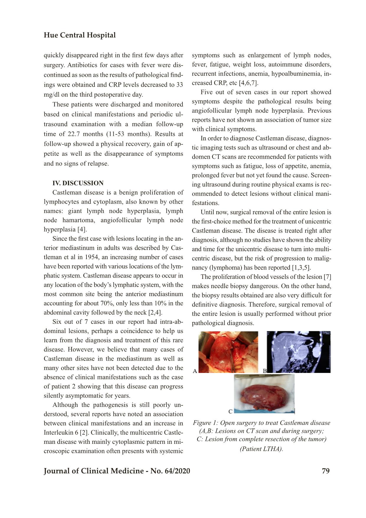
Figure 1: Open surgery to treat Castleman disease
(A,B: Lesions on CT scan and during surgery;
C: Lesion from complete resection of the tumor)
(Patient LTHA).
Bệnh viện Trung ương Huế
80 Journal of Clinical Medicine - No. 64/2020
All 7 of our cases were indicated for total surgi-
cal resection based on computed tomography evalu-
ation. During the operation we noted that the sur-
gery was relatively uneventful due to the relatively
clear limitation of the lesion, but care must be taken
to reveal and protect important blood vessels, which
in our cases are visceral artery, mesenteric artery
and carotid bundle. Lesions may be in one location
but may include multiple lymph nodes and need to
remove all of them.
It seems that lesions may be removed either by
open or laparoscopic surgery, even though lesions
locating adjacent to important blood vessels can
make laparoscopic surgery more dangerous. Lapa-
roscopic surgery is also helpful for us to locate the
lesion and select the appropriate abdominal route in
operation. Preoperative embolization by intravas-
cular intervention - in cases of extensive vascular
hyperplasia and in lesions at unfavorable locations
– to help a complete resection has been reported [7].
Figure 2: Laparoscopic surgery in the treatment of
found an appropriate answer to the clinical manifes-
tations of the two remaining cases although the path-
ological results of all 4 cases were Castleman disease
with angiofollicular lymph node hyperplasia.
Figure 3: Microscopic images of Castleman dis-
ease with angiofollicular lymph node hyperplasia
A. Hyperplasia of follicles in the lymph node.
B. “Lollipop” sign (head of arrow)
(Patient LTHA).
Many reports show that for unicentric Castleman
disease, surgical removal can be completely cured,
but there are cases of relapses after many years (1-7).
Therefore, it is necessary to monitor long-term for
cases of surgical removal.
Relapse or unresectable lesions or multicentric
disease can be treated with a variety of therapies
such as corticosteroid monotherapy or chemother-
apy. Other methods currently being applied, such as
interferon alpha, retinoic acid and anti-IL6 mono-
clonal antibodies, also show promising results.
V. CONCLUSIONS
Castleman disease is a rare disease in children
with a diverse clinical presentation characterized by
proliferation of lymph nodes with single or diffuse
lesions. Complete surgical removal is the treatment
of choice for unicentric Castleman disease.
Ultrasound and computed tomography are effec-
tive means of detecting the disease.
The outcome of surgery is favorable, but patients
need long-term follow-up to check for a relapse.
Castleman disease
(Patient TNTA).
The patients recovered well and there were no
complications. We noted a rapid disappearance of
symptoms in two cases ID 3 and 4 right in the first
few days of postoperation, especially the CRP level
of the third case decreased to 33 mg/dl on the third
postoperative day. It seems that the abrupt elimina-
tion of secreting chemicals from the tumors (such as
IL6) is the cause of this recovery but we have not
A B
REFERENCES
1. Dung TN, Vu KV. A cases serie of Castleman
disease. Y hoc thuc hanh. 2012 December; 855,
pp 129-131.
2. Kathryn M, Heather E, Andreana B. Intraab-
dominal mass and Iron deficiency anemia in a 15
year old boy: case report and literature review.
Journal of pediatric surgery. 2014; case reports
2, pp. 123-125.
Castleman disease in children: Diagnosis and treatment
Hue Central Hospital
Journal of Clinical Medicine - No. 64/2020 81
3. Keely B, Deborah P, Christine R, Shahab A.
Castleman disease: surgical cure in pediatric pa-
tients. Journal of pediatric surgery. 2009. 44, pp.
E5-E8.
4. Ke RZ, Hui MJ(2008). Mesenteric castleman
disease. Journal of pediatric surgery.2008; 44,
pp.1398-1400.
5. Idil RU, Zuhal A, Ibrahim K. Castleman dis-
ease: an usual diagnosis of a portal mass in an 8
year old boy. Journal of pediatric surgery, 2010;
46, pp. E9-E11.
6. Jan FS, Mats MH, et al. Minimal access surgery
in cactleman disease in a child, a case report.
Journal of pediatric surgery.2015; case reports 3,
pp. 289-291.
7. Shawn DS, Anand SL, Samuel AM. Preop-
erative embolization as an adjunct to the op-
erative management of mediastinal castleman
disease. Journal of pediatric surgery. 2003; 38
No9, pp. E43.
File đính kèm:
 castleman_disease_in_children_diagnosis_and_treatment.pdf

