Bài giảng Thông khí tư thế nằm sấp trong ards (Nhân 1 ca lâm sàng)
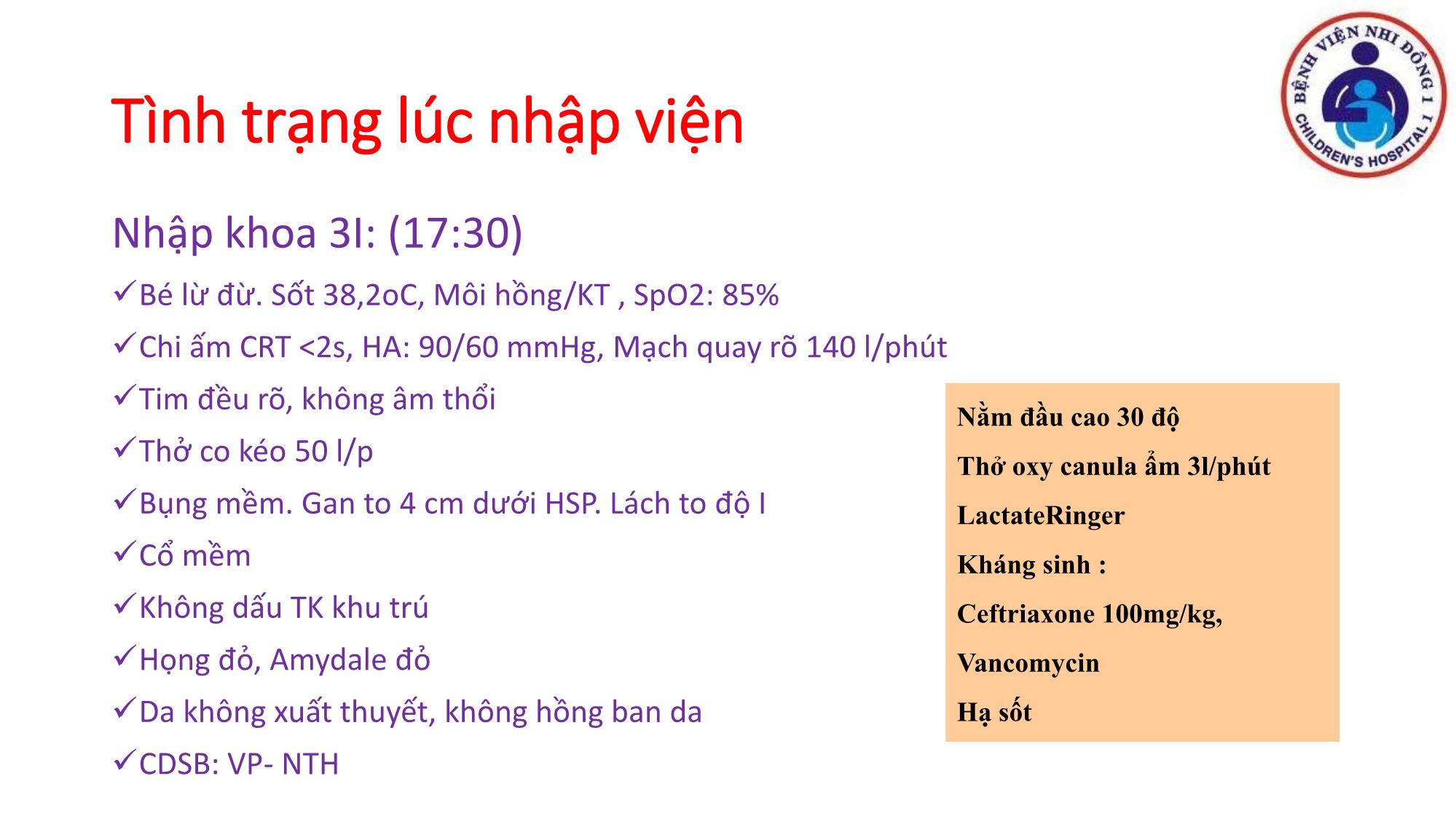
Tình trạng lúc nhập viện
Nhập khoa 3I: (17:30)
✓Bé lừ đừ. Sốt 38,2oC, Môi hồng/KT , SpO2: 85%
✓Chi ấm CRT <2s, HA: 90/60 mmHg, Mạch quay rõ 140 l/phút
✓Tim đều rõ, không âm thổi
✓Thở co kéo 50 l/p
✓Bụng mềm. Gan to 4 cm dưới HSP. Lách to độ I
✓Cổ mềm
✓Không dấu TK khu trú
✓Họng đỏ, Amydale đỏ
✓Da không xuất thuyết, không hồng ban da
✓CDSB: VP- NTH
Nằm đầu cao 30 độ
Thở oxy canula ẩm 3l/phút
LactateRinger
Kháng sinh :
Ceftriaxone 100mg/kg,
Vancomycin
Hạ sốtCận lâm sàng
WBC: 3.71 K/uL NEU: 1.30
(35%)
RBC : 4,63 M/uL Hb: 11.5 g/dl
Hct: 33.5%
PLT : 133 K/uL
PT: 11.8s
APTT: 30.6s
INR: 0.85
Fibrinogen: 3.69 g/l
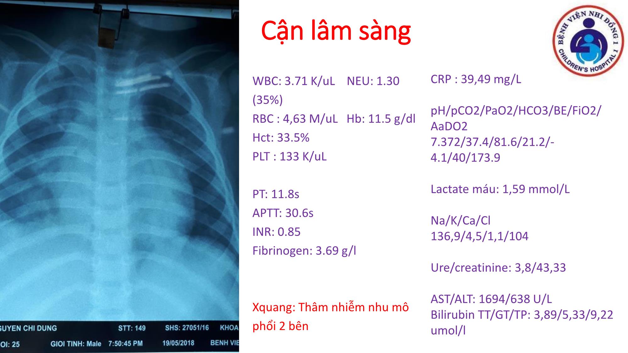
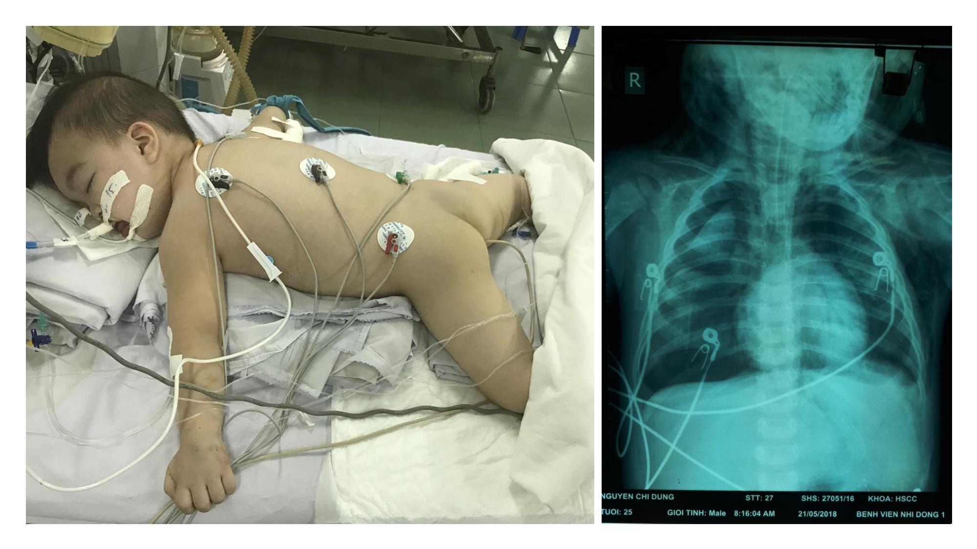
Xquang: Thâm nhiễm nhu mô
phổi 2 bên
CRP : 39,49 mg/L
pH/pCO2/PaO2/HCO3/BE/FiO2/
AaDO2
7.372/37.4/81.6/21.2/-
4.1/40/173.9
Lactate máu: 1,59 mmol/L
Na/K/Ca/Cl
136,9/4,5/1,1/104
Ure/creatinine: 3,8/43,33
AST/ALT: 1694/638 U/L
Bilirubin TT/GT/TP: 3,89/5,33/9,22
umol/l

Trang 1
Trang 2
Trang 3
Trang 4
Trang 5
Trang 6
Trang 7
Trang 8
Trang 9
Trang 10
Tải về để xem bản đầy đủ
Tóm tắt nội dung tài liệu: Bài giảng Thông khí tư thế nằm sấp trong ards (Nhân 1 ca lâm sàng)

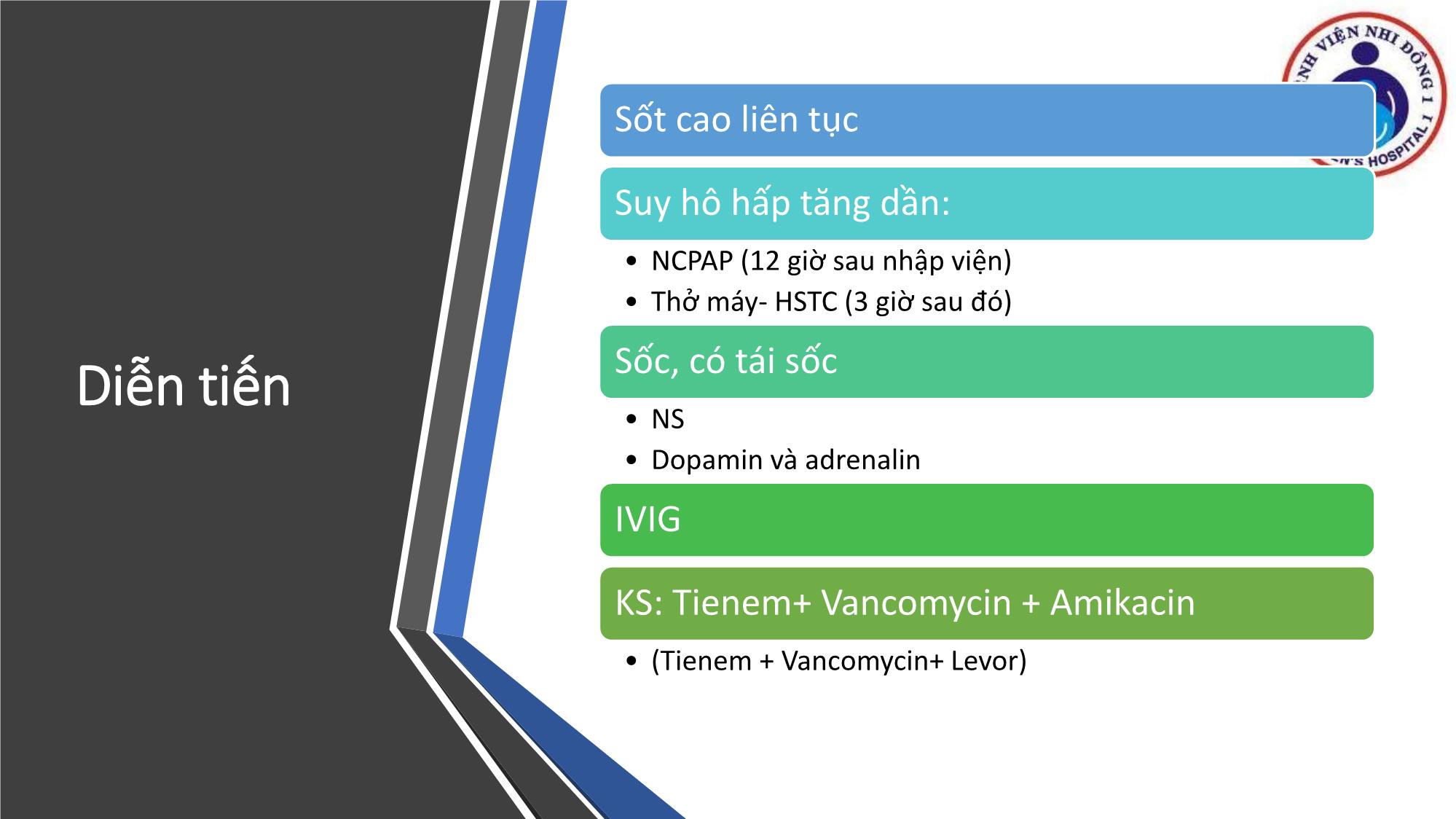
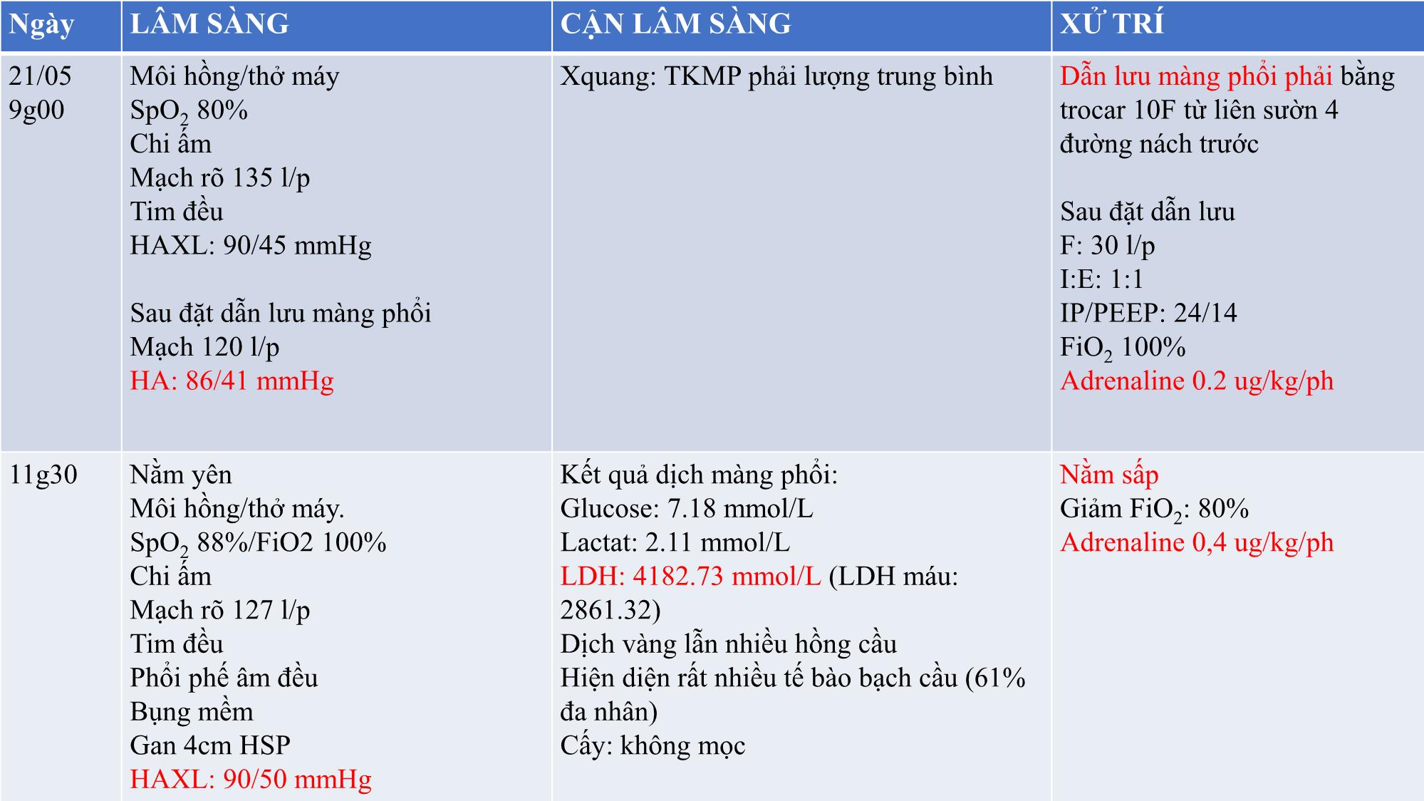
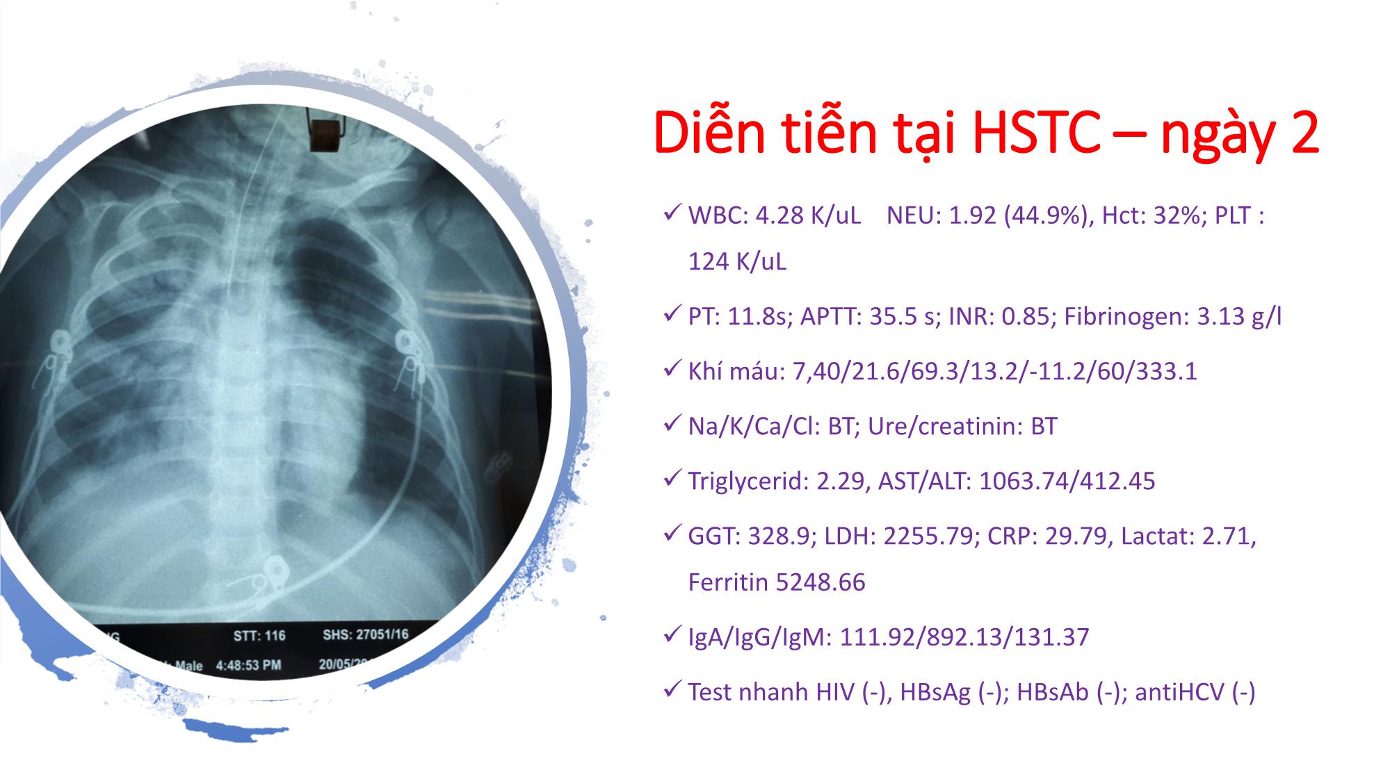
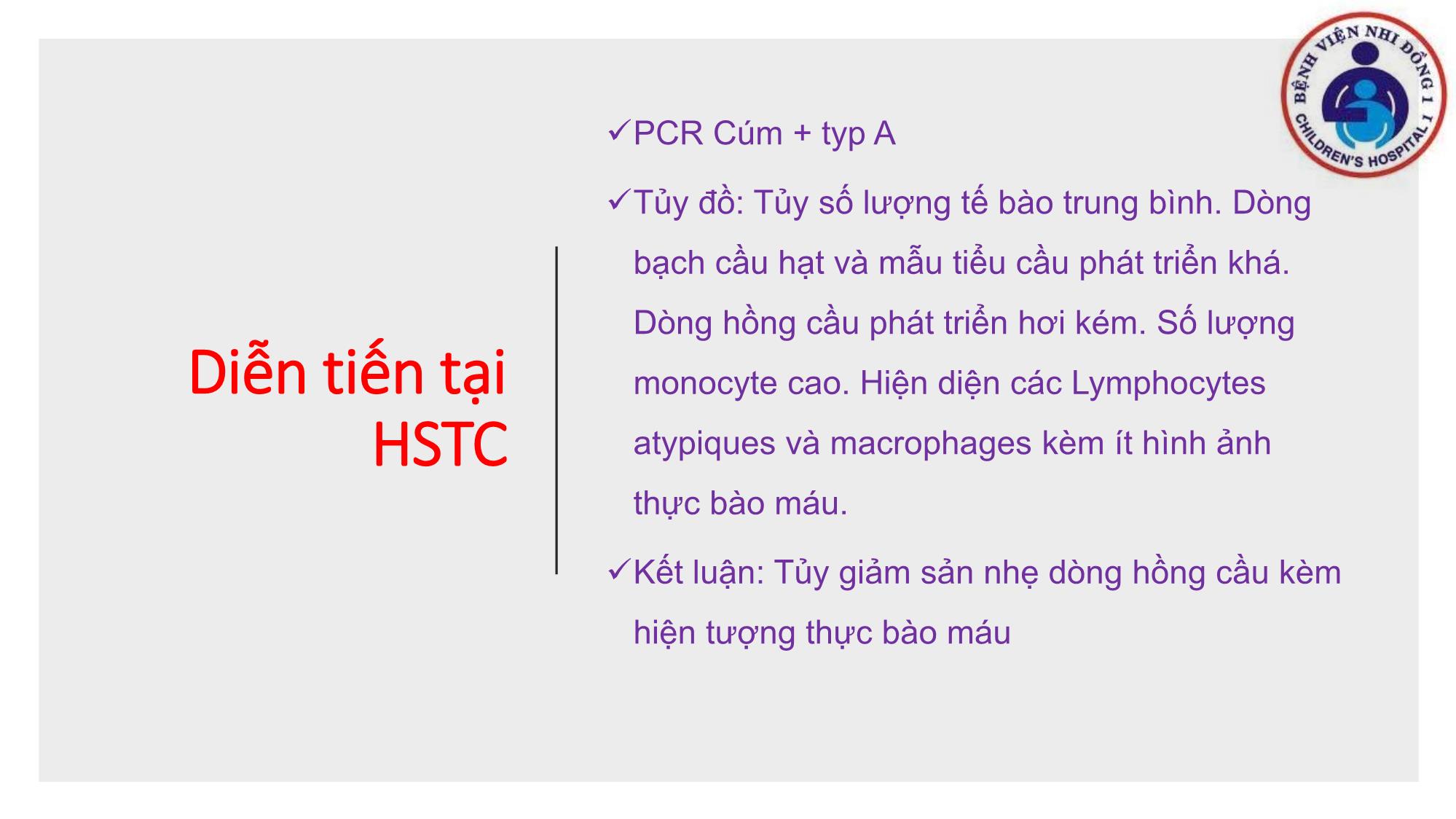
THÔNG KHÍ TƯ THẾ NẰM SẤP TRONG ARDS (Nhân 1 ca lâm sàng) Khoa HSTCCĐ Bv Nhi Đồng 1 HÀNH CHÁNH ✓Họ và tên : NGUYỄN CHÍ D ✓Tuổi : 30 tháng tuổi ✓Giới tính : Nam ✓Địa chỉ : Quận Bình Tân, TPHCM ✓Nhập viện: 17g30 ngày 19/05/2018 ✓Nhập viện vì sốt BỆNH SỬ Bệnh 6 ngày ✓Bé sốt cao 39 – 40oC, ho khan nhiều. ✓Không co giật, không lạnh run. Giảm chơi, ăn uống kém. Không ói,. Tiểu được, vàng trong, không lắt nhắt. ✓Tiêu phân vàng sệt, 1-2 lần/ngày; không đau bụng. => Nhập BV Nhi đồng 1 vì sốt cao sau điều trị Bs tư không giảm Tình trạng lúc nhập viện Nhập khoa 3I: (17:30) ✓Bé lừ đừ. Sốt 38,2oC, Môi hồng/KT , SpO2: 85% ✓Chi ấm CRT <2s, HA: 90/60 mmHg, Mạch quay rõ 140 l/phút ✓Tim đều rõ, không âm thổi ✓Thở co kéo 50 l/p ✓Bụng mềm. Gan to 4 cm dưới HSP. Lách to độ I ✓Cổ mềm ✓Không dấu TK khu trú ✓Họng đỏ, Amydale đỏ ✓Da không xuất thuyết, không hồng ban da ✓CDSB: VP- NTH Nằm đầu cao 30 độ Thở oxy canula ẩm 3l/phút LactateRinger Kháng sinh : Ceftriaxone 100mg/kg, Vancomycin Hạ sốt Cận lâm sàng WBC: 3.71 K/uL NEU: 1.30 (35%) RBC : 4,63 M/uL Hb: 11.5 g/dl Hct: 33.5% PLT : 133 K/uL PT: 11.8s APTT: 30.6s INR: 0.85 Fibrinogen: 3.69 g/l Xquang: Thâm nhiễm nhu mô phổi 2 bên CRP : 39,49 mg/L pH/pCO2/PaO2/HCO3/BE/FiO2/ AaDO2 7.372/37.4/81.6/21.2/- 4.1/40/173.9 Lactate máu: 1,59 mmol/L Na/K/Ca/Cl 136,9/4,5/1,1/104 Ure/creatinine: 3,8/43,33 AST/ALT: 1694/638 U/L Bilirubin TT/GT/TP: 3,89/5,33/9,22 umol/l Diễn tiến Sốt cao liên tục Suy hô hấp tăng dần: • NCPAP (12 giờ sau nhập viện) • Thở máy- HSTC (3 giờ sau đó) Sốc, có tái sốc • NS • Dopamin và adrenalin IVIG KS: Tienem+ Vancomycin + Amikacin • (Tienem + Vancomycin+ Levor) Ngày LÂM SÀNG CẬN LÂM SÀNG XỬ TRÍ 21/05 9g00 Môi hồng/thở máy SpO2 80% Chi ấm Mạch rõ 135 l/p Tim đều HAXL: 90/45 mmHg Sau đặt dẫn lưu màng phổi Mạch 120 l/p HA: 86/41 mmHg Xquang: TKMP phải lượng trung bình Dẫn lưu màng phổi phải bằng trocar 10F từ liên sườn 4 đường nách trước Sau đặt dẫn lưu F: 30 l/p I:E: 1:1 IP/PEEP: 24/14 FiO2 100% Adrenaline 0.2 ug/kg/ph 11g30 Nằm yên Môi hồng/thở máy. SpO2 88%/FiO2 100% Chi ấm Mạch rõ 127 l/p Tim đều Phổi phế âm đều Bụng mềm Gan 4cm HSP HAXL: 90/50 mmHg Kết quả dịch màng phổi: Glucose: 7.18 mmol/L Lactat: 2.11 mmol/L LDH: 4182.73 mmol/L (LDH máu: 2861.32) Dịch vàng lẫn nhiều hồng cầu Hiện diện rất nhiều tế bào bạch cầu (61% đa nhân) Cấy: không mọc Nằm sấp Giảm FiO2: 80% Adrenaline 0,4 ug/kg/ph Diễn tiễn tại HSTC – ngày 2 ✓WBC: 4.28 K/uL NEU: 1.92 (44.9%), Hct: 32%; PLT : 124 K/uL ✓ PT: 11.8s; APTT: 35.5 s; INR: 0.85; Fibrinogen: 3.13 g/l ✓ Khí máu: 7,40/21.6/69.3/13.2/-11.2/60/333.1 ✓Na/K/Ca/Cl: BT; Ure/creatinin: BT ✓ Triglycerid: 2.29, AST/ALT: 1063.74/412.45 ✓GGT: 328.9; LDH: 2255.79; CRP: 29.79, Lactat: 2.71, Ferritin 5248.66 ✓ IgA/IgG/IgM: 111.92/892.13/131.37 ✓ Test nhanh HIV (-), HBsAg (-); HBsAb (-); antiHCV (-) Diễn tiến tại HSTC ✓PCR Cúm + typ A ✓Tủy đồ: Tủy số lượng tế bào trung bình. Dòng bạch cầu hạt và mẫu tiểu cầu phát triển khá. Dòng hồng cầu phát triển hơi kém. Số lượng monocyte cao. Hiện diện các Lymphocytes atypiques và macrophages kèm ít hình ảnh thực bào máu. ✓Kết luận: Tủy giảm sản nhẹ dòng hồng cầu kèm hiện tượng thực bào máu PARDS Adult Respiratory Distress Syndrome (Ashbaugh-1967) Acute Respiratory Distress Syndrome (1991?) Acute Respiratory Distress Syndrome (AECC-1994) Ped Acute Respiratory Distress Syndrome (PARDS- 2015) Acute Respiratory Distress Syndrome (Berlin-2012) 125 dependent and less positive in dependent regions [51,52] . The net effect of prone positioning is not only the increase of regional inflation distribution in dorsal regions and decrease in ventral regions respectively, but intrapleural pressure, transpulmonary pressure and regional inflat i on distribution become more homo geneous throughout the lung (Figure 2) [53] . It was early suggested that this could be explained by the reversal of lung weight gradients, the direct transmission of the weight of the heart to subjacent regions, direct transmission of the weight of abdominal contents to caudal regions of the dorsal lung and/or regional mechanical properties and shape of the chest wall and lung [54] . In addition, although in healthy lung pulmonary perfusion is distributed along a ventral-dorsal gradient and progressively increases down the lung, data suggest that in the diseased lung, blood flow is being diverted toward the nondependent regions. This is caused by several mechanisms including hypoxic vasoconstriction, vessel obliteration and extrinsic vessel compression [5557] . Also, human and animal studies have shown that in the conversion from the supine to prone ventilation, pulmonary blood flow in dorsal regions of the lung is maintained unmodified and prevalent in lung dorsal areas (Figure 2) [46,53,5863] . Besides, in patients with ARDS, the increased lung weight due to serious inflammation and pulmonary edema would act also as hypergravity to squeeze the blood flow as well as ventilation out of the dependent area to the nondependent region [16,64] . Thus, the reduction in intrapulmonary shunt and the increase in oxygenation observed in patients with ARDS who are turned in the prone position mainly results from better ventilated well-perfused lung areas with dorsal recruitment being in parallel greater than ventral de-recruitment (Figure 2) [36,51,53] . Animal data had early suggested that during prone position homogeneity of ventilation increases V/Q as well as the correlation between regional ventil- ation and perfusion [65] . Very important is the finding that prone position, when combined, is followed by an improved and/or a more sustained response to recruiting maneuvers [50,6668] . Albert et al [47] in their study determined the fraction of lung that might be subjected to the weight of the heart when patients are in the supine vs the prone position. The study included only nonARDS patients, but it was found that turning patients to the prone position eliminates the compressive force of the heart on dorsal lung regions redirecting it to only a small portion of the ventral lung regions (Figure 3). This is in agreement with the results of previous studies. In a study conducted by our group it was shown that ARDS patients with congestive heart failure and cardiomegaly after being turned to the prone positioning exhibited a significant, rapid, and persistent improvement in oxygenation. This improvement could be partly due to the decompression of the left lower lobe by the enlarged heart [69] . Wiener et al [70] had early found that patients with cardiomegaly exhibited reduced left mid- and lower zone ventilation in the supine but not in the prone position. May 4, 2016|Volume 5|Issue 2|WJCCM|www.wjgnet.com Prone positionSupine position Minutes Dorsal Time Ventral Shape matchingIsolated lung No gravity Gravity Gravity Shape matching - B lo o d f lo w + A B C D E F Figure 2 A summary showing the sequential effects of prone position on acute respiratory distress syndrome diseased lung. A: Original shape of the isolated lung; the dorsal side is bigger than the ventral one (no gravity); B: The result of shape matching: alveolar units have bigger size ventrally and smaller size dorsally (no gravity); C: The additive effect of gravity on ventilation and perfusion: blood flow is being diverted toward dependent regions, while dependent pulmonary units close; D: Immediately after turning to the prone position, pulmonary blood flow in dorsal regions of the lung is maintained unmodifie; E: Dorsal lung recruitment follows (greater than ventral de-recruitment), gravitational forces compress the ventral region, but this effect is damped by regional expansion due to shape matching; F: Transpulmonary pressure and regional infla t ion di st ribut ion become mo r e homo geneous thr oughout the lung resul ting fina l l y t o bet t er oxygenat i on. Koulouras V et al . Pathophysiology of prone position in ARDS Chỉ định ✓Indication ▪ Severe ARDS • PaO2/FiO2 < 100 mmHg ▪ Fail to improve with conventional ventilatory strategies Chống chỉ định Chống chỉ định tương đối Biến chứng PRONE PROCEDURE ✓Timing of initiation: 12-24 hours after supine ▪ Early initiation of prone ventilation was most effective, ▪ Collapsed lung units are likely to be opened (ie, recruited) most easily during the acute exudative phase of ARDS ✓Prone ventilation early (up to 36 hours) Patients ✓ARDS: American–European Consensus Conference criteria ▪ Endotracheal intubation and mechanical ventilation for ARDS for less than 36 hours; and ▪ Severe ARDS: • Pao2/Fio2 <150 mm Hg, Fio2 of ≥0.6, PEEP ≥5 cm • Vt: 6 ml/kg. ▪ The criteria were confirmed after 12 to 24 hours ✓Question:How does prone positioning compare with supine positioning for ventilation in adults with severe acute respiratory failure? Clinical Answer: ✓In people with severe acute respiratory failure, prone positioning during ventilation, rather than supine positioning, reduces mortality in specific clinical situations, but is associated with a higher risk of adverse events. AnnalsATS Volume 14 Supplement 4|October 2017 ✓102 BN/8017 , 2 tuần- 18 tuổi ✓Nằm sấp sống 80% ✓Nằm ngữa 86%. Kết luận ✓PARDS tỉ lệ tử vong cao, có nhiều phương tiện giúp giải quyết vấn đề giảm oxy máu. ✓Nằm sấp là một lựa chọn đang được ủng hộ. ✓Khi có chỉ định nằm sấp thì nên được thực hiện sớm. ✓Theo dõi sát, đánh giá hiệu quả và thực hiện nằm sấp đủ thời gian. ✓Theo dõi sát những biến chứng trong quá trình sằm sấp. * Ths. Bs. Bùi Thanh Liêm - Bộ môn nhi - ĐHYD TPHCM - Khoa HSTCCĐ BV Nhi Đồng 1 - Email: liem.bui@ump.edu.vn - Cellphone: +84 938165083
File đính kèm:
 bai_giang_thong_khi_tu_the_nam_sap_trong_ards_nhan_1_ca_lam.pdf

